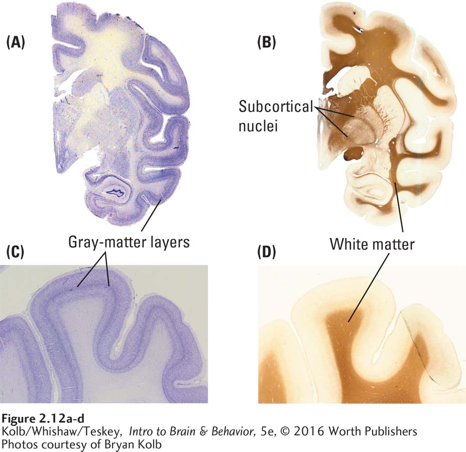
FIGURE 2- 12 Cortical Layers and Glia Brain sections from the left hemisphere of a monkey (midline is to the left in each image), viewed through a microscope. Cells are stained with (A and C) a selective cell body stain for neurons (gray matter) and (B and D) a selective fiber stain for insulating glial cells, or myelin (white matter). The images reveal very different views of the brain at the macro (A and B) and microscopic (C and D) levels.
Photos courtesy of Bryan Kolb