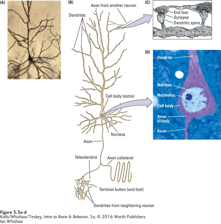
FIGURE 3- 3 Major Parts of a Neuron (A) Typical neuron Golgi- stained to reveal its dendrites and cell body. (B) The neuron’s basic structures identified. (C) An electron micrograph captures the synapse between an axon from another neuron and a dendritic spine. (D) High- power light microscopic view inside the cell body. Note the axon hillock at the junction of the soma and axon.
Ian Whishaw