Specialized features are found in some prokaryotes
As they evolved, some prokaryotes developed specialized structures that gave them a selective advantage: cells with these structures were better able to survive and reproduce in particular environments than cells lacking them.
CELL WALLS Most prokaryotes have a cell wall located outside the cell membrane. The rigid wall supports the cell and determines its shape. The cell walls of most bacteria, but not archaea, contain peptidoglycan, a polymer of amino sugars that is linked at regular intervals to short peptides. Cross-
Enclosing the cell wall in some bacteria is a slimy layer composed mostly of polysaccharides and referred to as a capsule. In some cases these capsules protect the bacteria from attack by white blood cells in the animals they infect. Capsules also help keep the cells from drying out, and sometimes they help bacteria attach to other cells.
INTERNAL MEMBRANES Some groups of bacteria—
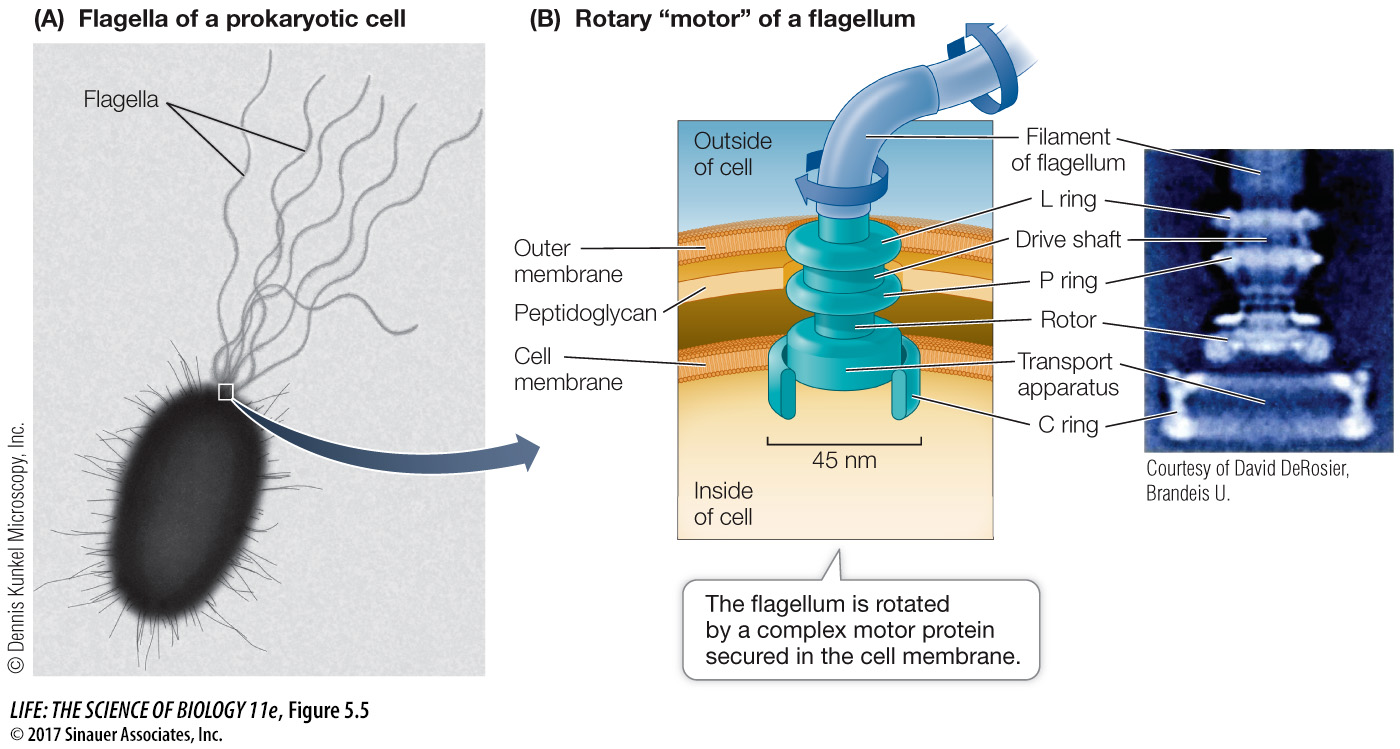
FLAGELLA AND PILI Some prokaryotes swim by using appendages called flagella, which sometimes look like tiny corkscrews (Figure 5.5A). In bacteria, the filament of the flagellum is made of a protein called flagellin. (As you will see in Key Concept 5.3, the flagella of eukaryotes are quite different in structure, but similar in function.) A complex motor protein spins the bacterial flagellum on its axis like a propeller, driving the cell along. The motor protein is anchored to the cell membrane and, in some bacteria, to the outer membrane of the cell wall (Figure 5.5B). We know that the flagella cause the motion of cells because if they are removed, the cells do not move.
Pili are structures made of protein that project from the surfaces of some types of bacterial cells. These hairlike structures are shorter than flagella and are used for adherence. Conjugative pili (sex pili) help bacteria join to one another to exchange genetic material. Fimbriae are composed of the same proteins as pili but are shorter, and help cells adhere to surfaces such as animal cells, for food and protection.
CYTOSKELETON The cytoskeleton is the collective name for protein filaments that play roles in cell division, cell movement, and in maintaining the shapes of cells. One such protein forms a ring structure that constricts during cell division, whereas another forms helical structures that extend down the lengths of rod-