Organelles can be studied by microscopy or isolated for chemical analysis
Cell organelles and structures were first detected by light and then by electron microscopy. The functions of the organelles could sometimes be inferred by observations and experiments, leading, for example, to the hypothesis (later confirmed) that the nucleus contained the genetic material. Later, the use of stains targeted to specific macromolecules allowed cell biologists to determine the chemical compositions of organelles.
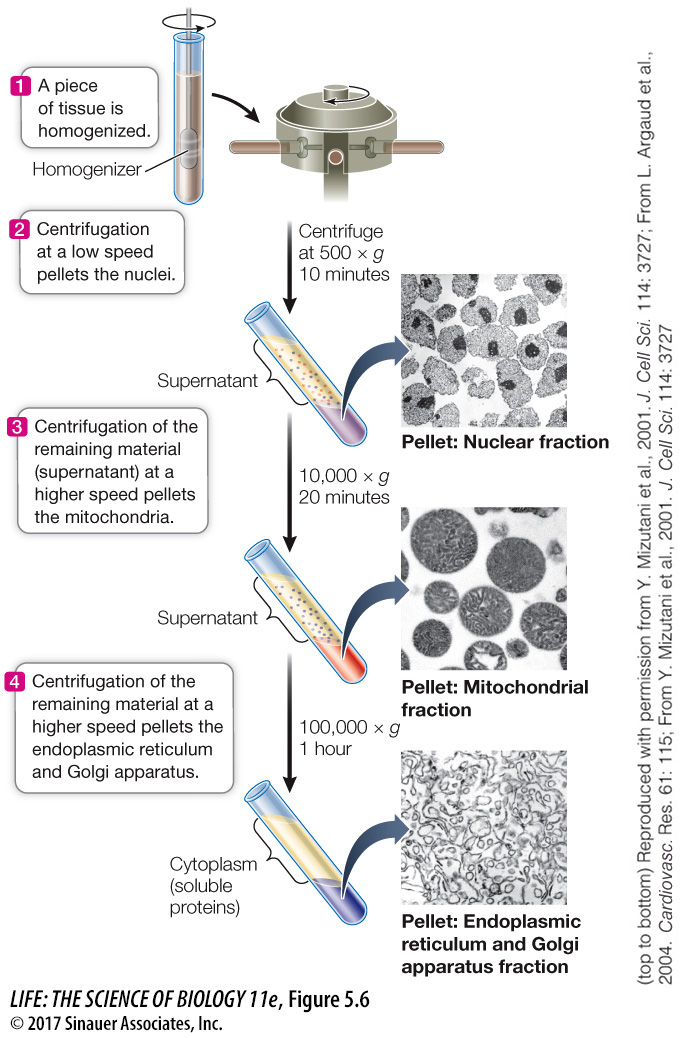
Another way to analyze cells is to take them apart in a process called differential centrifugation, or cell fractionation. This process permits cell organelles and other cytoplasmic structures to be separated from each other and examined using chemical methods. Cell fractionation begins with the destruction of the cell membrane, which allows the cytoplasmic components to flow out into a test tube. The various organelles can then be separated from one another on the basis of size or density (Figure 5.6). Biochemical analyses can then be done on the isolated organelles. Microscopy and cell fractionation have complemented each other, giving us a more complete picture of the composition and function of each organelle and structure.
research tools
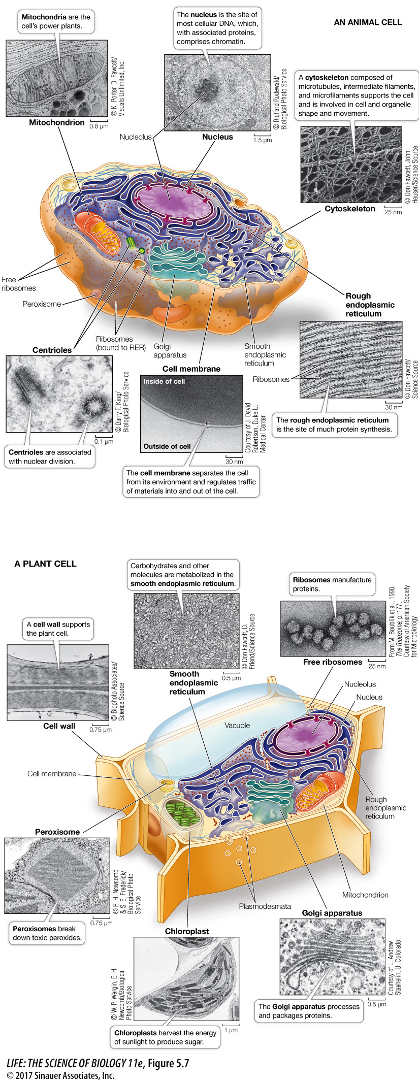
Microscopy of eukaryotic cells has revealed that many of the organelles are similar in appearance in each cell type (Figure 5.7). By comparing Figures 5.7 and 5.4 you can see some of the prominent differences between eukaryotic cells and prokaryotic cells.
Media Clip 5.1 The Inner Life of a Cell