The spindle begins to form during prophase
During interphase, only the nuclear envelope, the nucleoli (see Key Concept 5.3), and a barely discernible tangle of chromatin are visible under the light microscope. The appearance of the nucleus changes as the cell enters prophase. At this stage, most of the cohesin that has held the two products of DNA replication together since S phase is removed, so the individual chromatids become visible. They are still held together by a small amount of cohesin at the centromere. Late in prophase, specialized structures called kinetochores develop in the centromere region, one on each chromatid. These structures will be important for chromosome movement.
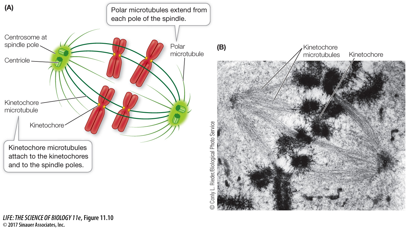
Each of the two centrosomes, now on opposite sides of the nucleus, serves as a mitotic center, or pole, toward which the chromosomes will move (Figure 11.10A). During prophase and prometaphase, microtubules form between the poles and the chromosomes to make up the spindle. The spindle serves as a structure to which the chromosomes attach and as a framework keeping the two poles apart. Each half of the spindle develops as tubulin dimers aggregate from around the centrioles and form long fibers that extend into the middle region of the cell. The microtubules are initially unstable, constantly forming and falling apart, until they contact kinetochores or microtubules from the other half-
Activity 11.2 The Mitotic Spindle
There are two groups of microtubules in the spindle:
Polar microtubules form the framework of the spindle and run from one pole to the other.
Kinetochore microtubules, which form later, attach to the kinetochores on the chromosomes. The two sister chromatids in each chromosome pair become attached to kinetochore microtubules in opposite halves of the spindle (Figure 11.10B). This ensures that the two chromatids will eventually move to opposite poles.
Movement of the chromatids achieves the central goal of mitosis. It accomplishes the segregation of the genetic material that must occur before the cell can divide and complete the cell cycle. Prophase prepares for this movement, and the actual segregation takes place in the next three phases of mitosis.