Four key features define DNA structure
Four features summarize the molecular architecture of the DNA molecule (Figure 13.6B):
DNA is a double-
stranded helix , with a sugar–phosphate backbone on the outside and base pairs lined up on the inside. DNA is usually a right-
handed helix . If you curl the fingers of your right hand and point your thumb upward, the curve of the helix follows the direction of your fingers, and it winds upward in the direction of your thumb. (See Figure 3.8, of right-versus left- handed helices.) DNA is antiparallel (the two strands run in opposite directions).
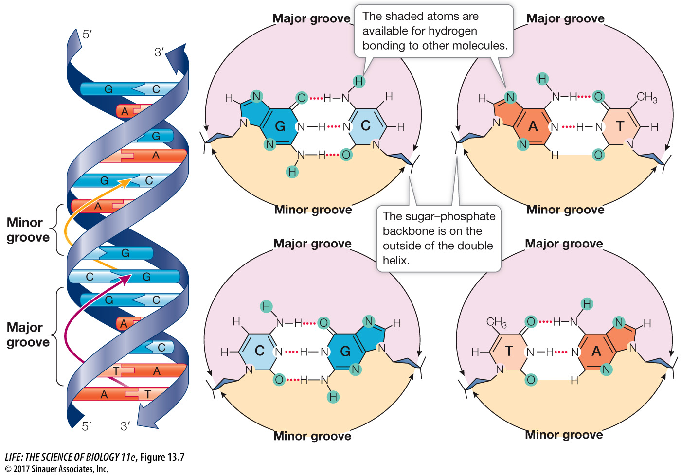
DNA has major and minor grooves in which the outer edges of the nitrogenous bases are exposed.
THE HELIX The sugar–
Hydrogen bonding between specifically paired bases. Consistent with Chargaff’s rule, adenine (A) pairs with thymine (T) by forming two hydrogen bonds, and guanine (G) pairs with cytosine (C) by forming three hydrogen bonds:
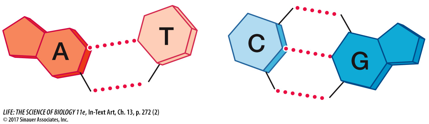
Every base pair consists of one purine (A or G) and one pyrimidine (T or C). This pattern is known as complementary base pairing.
van der Waals forces between adjacent bases on the same strand. When the base rings come near one another, they tend to stack like poker chips because of these weak attractions.
ANTIPARALLEL STRANDS The backbone of each DNA strand contains repeating units of the five-
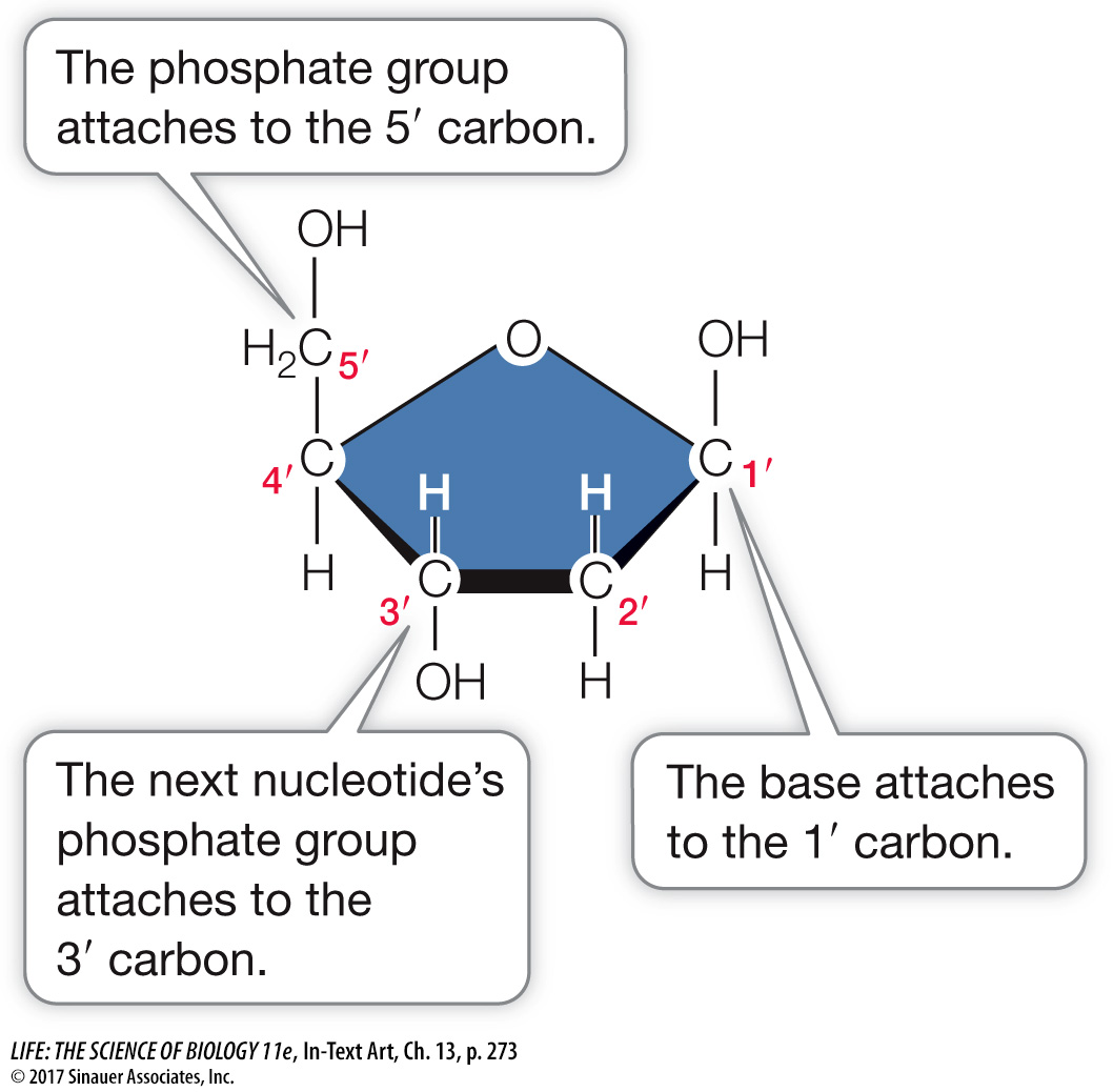
The number followed by a prime (′) designates the position of a carbon atom in this sugar molecule. In the sugar–
Thus the two ends of a polynucleotide chain differ. At one end of a chain is a free (not connected to another nucleotide) 5′ phosphate group (—OPO3–); this is called the 5′ end. At the other end is a free 3′ hydroxyl group (—OH); this is called the 3′ end. In a DNA double helix, the 5′ end of one strand is paired with the 3′ end of the other strand, and vice versa. In other words, if you drew an arrow for each strand running from 5′ to 3′, the arrows would point in opposite directions (see also Figure 4.4A).
BASE EXPOSURE IN THE GROOVES Look back at Figure 13.6B and note the major and minor grooves in the helix. These grooves exist because the backbones of the two strands are closer together on one side of the double helix (forming the minor groove) than on the other side (forming the major groove). Figure 13.7 shows a “top-