DNA from one type of bacterium genetically transforms another type
In the 1920s, English physician Frederick Griffith was studying the bacterium Streptococcus pneumoniae, or pneumococcus, one of the agents that cause pneumonia in humans (Figure 13.1). He was trying to develop a vaccine against this devastating illness (antibiotics had not yet been discovered). Griffith was working with two strains of pneumococcus:
Cells of the S strain produced colonies that looked smooth (S). Covered by a polysaccharide capsule, these cells were protected from attack by a host’s immune system. When S cells were injected into mice, they reproduced and caused pneumonia (the strain was virulent).
Cells of the R strain produced colonies that looked rough (R), lacked the protective capsule, and were not virulent.
experiment
Figure 13.1 Genetic Transformation
Original Paper: Griffith, F. 1928. The significance of pneumococcal types. Journal of Hygiene 27: 113–
Griffith’s experiments demonstrated that something in the virulent S strain of pneumococcus could transform nonvirulent R strain bacteria into a lethal form, even when the S strain bacteria had been killed by high temperatures.
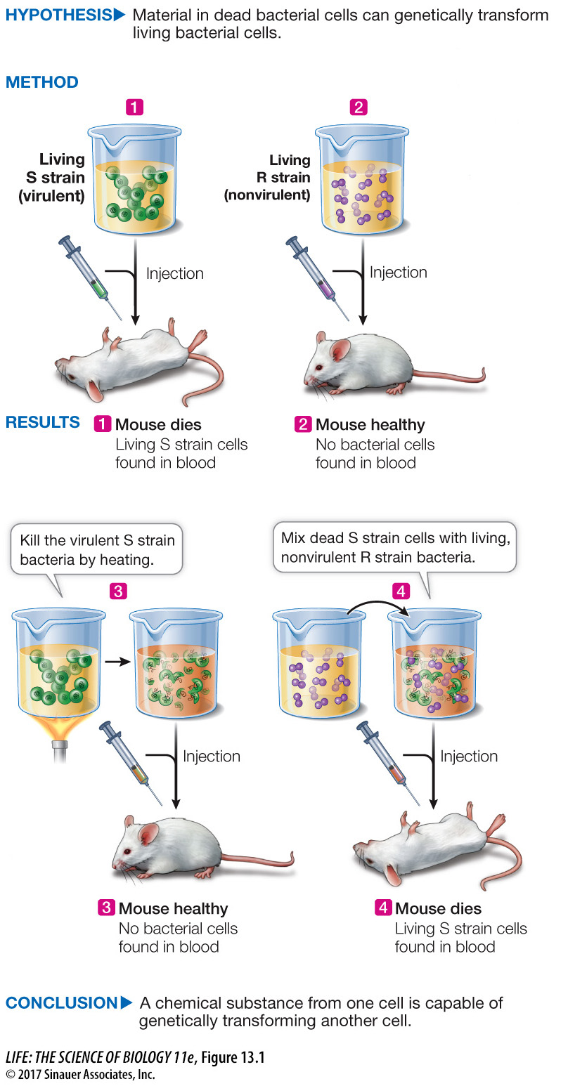
When Griffith injected mice with heat-
Did this transformation of the bacteria need to occur in the mouse’s body? No. The same transformation could be achieved in a test tube by mixing living R cells with heat-
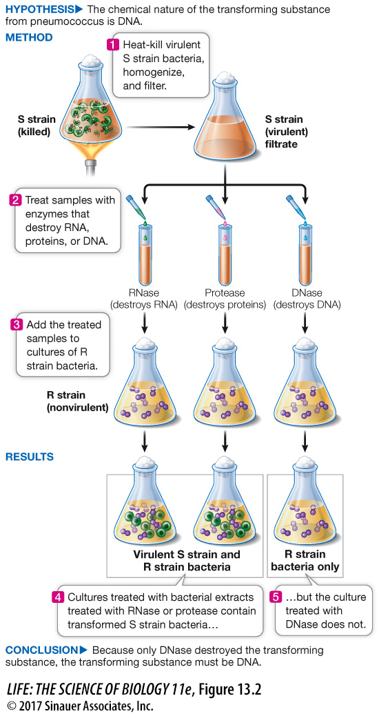
Oswald Avery and his colleagues at what is now The Rockefeller University identified the substance causing bacterial transformation in two ways:
Eliminating other possibilities. Cell-
free extracts containing the transforming substance were treated with enzymes that destroyed candidates for the genetic material, such as proteins, RNA, and DNA. When the treated samples were tested, the ones treated with RNase and protease (which destroy RNA and proteins, respectively) were still able to transform R- type bacteria into the S- type. But the transforming activity was lost in the extract treated with DNase (which destroys DNA) (Figure 13.2). Positive experiment. The researchers isolated virtually pure DNA from a cell-
free extract containing the transforming substance. The DNA alone caused bacterial transformation.
experiment
Figure 13.2 Genetic Transformation by DNA
Original Paper: Avery, O. T., C. M. MacLeod and M. McCarty. 1944. Studies on the chemical nature of the substance inducing transformation of the pneumococcal types. Journal of Experimental Medicine 79: 137–
Experiments by Avery and his colleagues showed that DNA from the virulent S strain of pneumococcus was responsible for the transformation in Griffith’s experiments (see Figure 13.1).
We now know that the gene for the enzyme that catalyzes the synthesis of the polysaccharide capsule, which makes the bacterial colony look “smooth,” was transferred into the R cells during transformation.