Viral infection experiments confirmed that DNA is the genetic material
Even with the bacterial transformation experiments, many biologists were still not convinced that DNA is the genetic material. One problem was that DNA, being made up of only four nucleotides (see Key Concept 4.1), seemed too uniform a substance to be able to confer instructions for all the functions and variety of life. The possibility remained that proteins, with all their chemical and structural diversity (20 amino acids), fulfilled that role. Experiments with a virus were designed to distinguish between these alternatives.
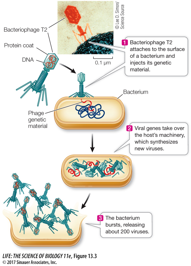
Alfred Hershey and Martha Chase of the Cold Spring Harbor Laboratory in New York studied bacteriophage T2 (phage T2), which infects the bacterium Escherichia coli. T2 phage consists of a DNA core packed inside a protein coat (Figure 13.3). When it attacks a bacterium, part (but not all) of the virus enters the bacterial cell. About 20 minutes later, the cell bursts, releasing dozens of particles that are virtually identical to the infecting virus particle. Clearly the virus is somehow able to hijack the host cell’s molecular machinery and convert it into a viral replication machine. Hershey and Chase set out to determine what part of the virus—
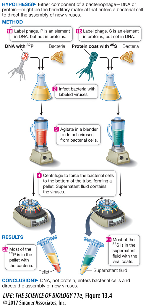
Proteins were labeled with radioactive sulfur. Proteins contain some sulfur (in the amino acids cysteine and methionine), but DNA does not. Sulfur has a radioactive isotope, 35S. Hershey and Chase grew bacteriophage T2 in a bacterial culture in the presence of 35S, so the proteins of the resulting viruses were labeled with (contained) the radioisotope.
DNA was labeled with radioactive phosphorus. DNA contains a lot of phosphorus (in the deoxyribose–
phosphate backbone— see Figure 4.4), whereas proteins contain little or none. Phosphorus also has a radioisotope, 32P. The researchers grew another batch of T2 in a bacterial culture in the presence of 32P, thus labeling the viral DNA with 32P.
Hershey and Chase used these radioactively labeled viruses in their experiments (Figure 13.4). In one experiment, they allowed 32P-
experiment
Figure 13.4 The Hershey–
Original Paper: Hershey, A. D. and M. Chase. 1952. Independent functions of viral protein and nucleic acid in growth of bacteriophage. The Journal of General Physiology 36: 39–
When Hershey and Chase infected bacterial cells with radioactively labeled T2 bacteriophage, only labeled DNA was found in the bacteria. The infected cells were agitated to remove the viral coats from the bacteria and were then centrifuged to pellet the bacteria. The labeled protein remained in the supernatant. This showed that DNA, not protein, is the genetic material.
A work with the data exercise that accompanies this figure may be assigned in LaunchPad.
Animation 13.1 The Hershey–