Gel electrophoresis separates DNA fragments
Restriction enzyme digestion is used to manipulate DNA in the laboratory so that mutations can be identified and analyzed. After a laboratory sample of DNA has been cut with one or more restriction enzymes, the DNA is in fragments, which must be separated to identify (map) where the cuts were made. Because the recognition sequence does not occur at regular intervals, the fragments are not all the same size, and these size differences can be used to separate the fragments from one another. Separating the fragments is necessary to determine the number and molecular sizes (in base pairs) of the fragments produced, or to identify and purify an individual fragment for further analysis or for use in an experiment.
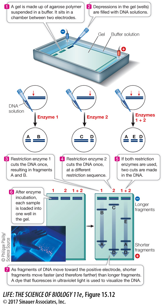
Gel electrophoresis is a common and convenient technique for separating or purifying DNA fragments. Samples containing the fragments are placed in wells at one end of a semisolid gel (usually made of agarose or polyacrylamide), and an electric field is applied to the gel (Figure 15.12). Because of its phosphate groups, DNA is negatively charged at neutral pH; therefore, because opposite charges attract, the DNA fragments move through the gel toward the positive end of the field. Because the spaces between the polymers of the gel are small, small DNA molecules can move through the gel faster than larger ones. Thus DNA fragments of different sizes separate from one another, forming bands that can be detected with a dye. This provides three types of information:
The number of fragments. The number of fragments produced by digestion of a DNA sample with a given restriction enzyme depends on how many times that enzyme’s recognition sequence occurs in the sample. Thus gel electrophoresis can provide some information about the presence of specific DNA sequences (the restriction sites) in the DNA sample.
The sizes of the fragments. DNA fragments of known size (size markers) are often placed in one well of the gel to provide a standard for comparison. The size markers are used to determine the sizes of the DNA fragments in samples in the other wells. By comparing the fragment sizes obtained with two or more restriction enzymes, the locations of their recognition sites relative to one another can be worked out (mapped).
The relative abundance of a fragment. In many experiments, the investigator is interested in how much DNA is present. The relative intensity of a band produced by a specific fragment can indicate the amount of that fragment.
research tools
Figure 15.12 Separating Fragments of DNA by Gel Electrophoresis A mixture of DNA fragments is placed in a gel, and an electric field is applied across the gel. The negatively charged DNA moves toward the positive end of the field, with smaller molecules moving faster than larger ones. After minutes to hours for separation, the electric power is shut off and the separated fragments can be analyzed.
Animation 15.1 Gel Electrophoresis