The small size of prokaryotes has hindered our study of their evolutionary relationships
Until about 300 years ago, nobody had even seen an individual prokaryote. Most prokaryotes remained invisible to humans until the invention of the first simple microscope. Prokaryotes are so small, however, that even the best light microscopes don’t reveal much about them. It took advanced microscopic equipment and modern molecular techniques to open up the microbial world. (Microscopic organisms—
Before DNA sequencing became practical, taxonomists based prokaryote classification on observable phenotypic characters such as shape, color, motility, nutritional requirements, and sensitivity to antibiotics. One of the characters most widely used to classify prokaryotes is the structure of their cell walls.
The cell walls of almost all bacteria contain peptidoglycan, a cross-
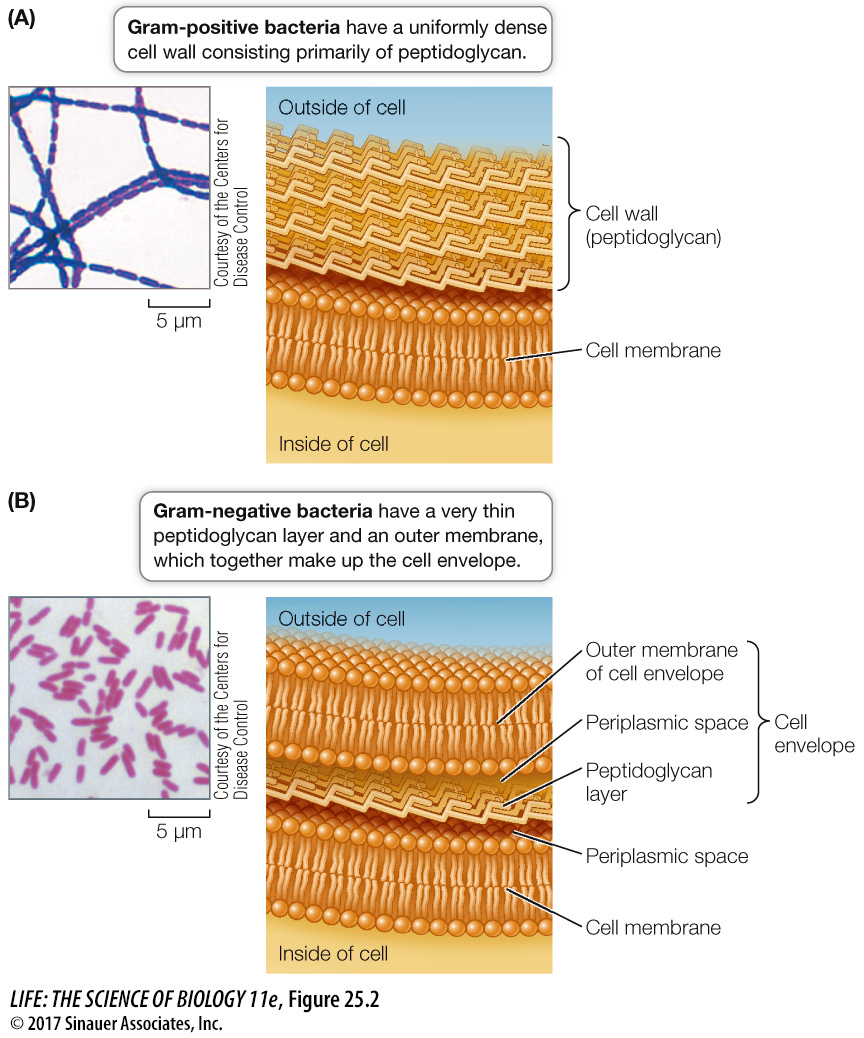
The Gram stain is a technique that can be used to separate most types of bacteria into two distinct groups. A smear of bacterial cells on a microscope slide is soaked in a violet dye and treated with iodine; it is then washed with alcohol and counterstained with a red dye called safranin. Gram-
A Gram-
negative cell wall usually has a thin peptidoglycan layer, which is surrounded by a second, outer membrane quite distinct in chemical makeup from the cell membrane (see Figure 25.2B). Together the cell wall and the outer membrane are called the cell envelope. The space between the cell membrane and the outer membrane (known as the periplasmic space) contains proteins that are important in digesting some materials, transporting others, and detecting chemical gradients in the environment.A Gram-
positive cell wall usually has about five times as much peptidoglycan as a Gram-negative cell wall. Its thick peptidoglycan layer is a meshwork that may serve some of the same purposes as the periplasmic space of the Gram- negative cell envelope.
Activity 25.1 Gram Stain and Bacteria
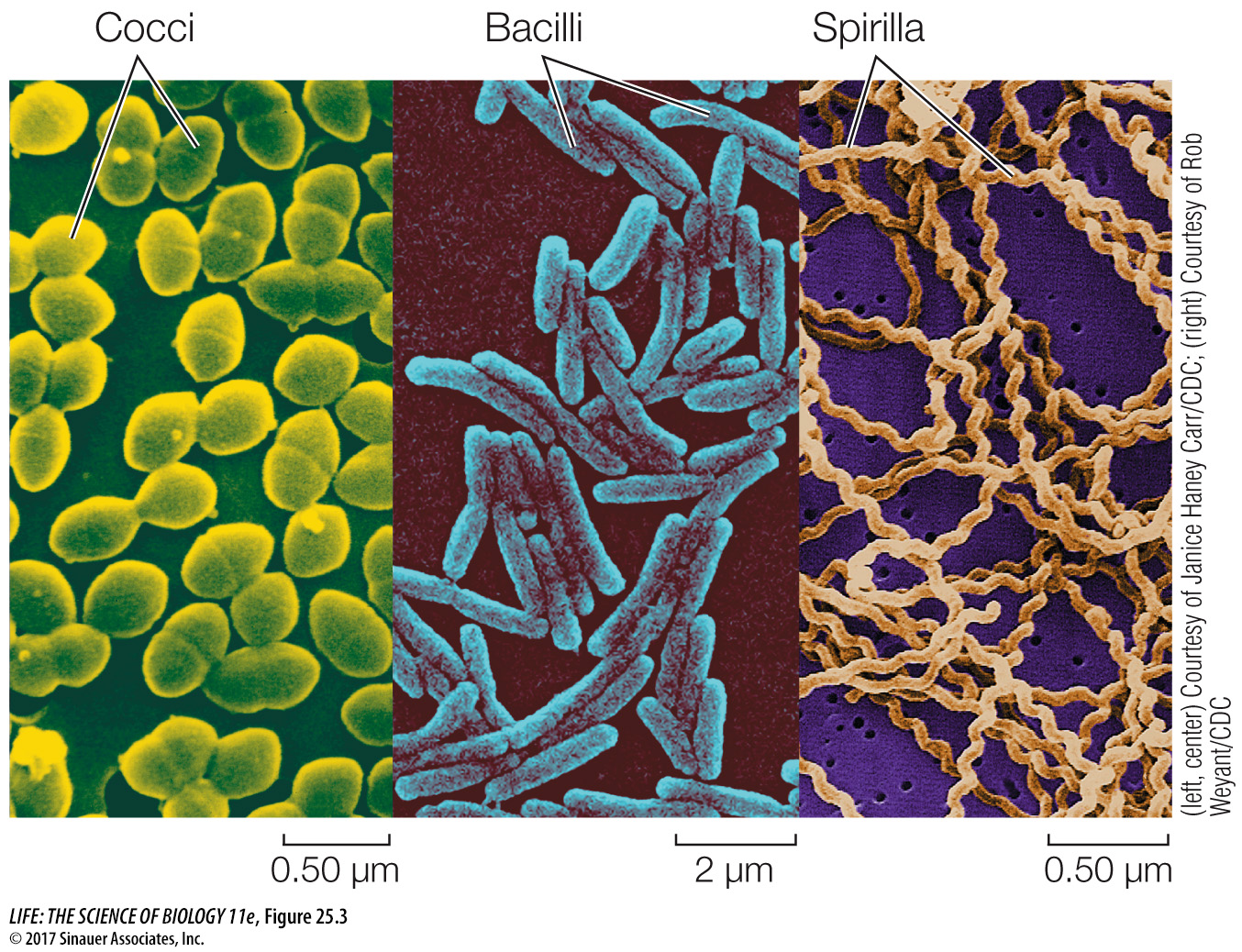
Shape is another phenotypic characteristic that is useful for the basic identification of bacteria. The three most common shapes are spheres, rods, and spiral forms (Figure 25.3). Many bacterial names are based on these shapes. A spherical bacterium is called a coccus (plural cocci). Cocci may live singly or may associate in two-
Less is known about the shapes of prokaryotic archaea because many of these organisms have never been seen. Many prokaryotic archaea are known only from samples of DNA from the environment. However, the species whose morphologies are known include cocci, bacilli, and even triangular and square species. Some flattened species grow on surfaces, arranged like sheets of postage stamps.