Excavates began to diversify about 1.5 billion years ago
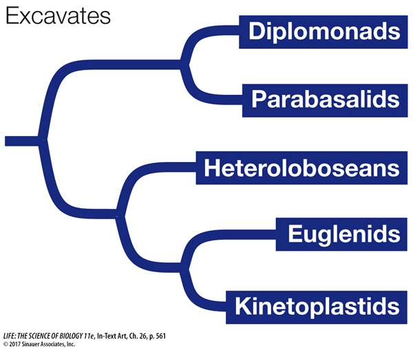
The excavates include a number of diverse groups that began to split from one another soon after the origin of eukaryotes. Several groups of excavates lack mitochondria, an absence that once led to the view that these groups might represent early-
DIPLOMONADS AND PARABASALIDS The diplomonads and the parabasalids are unicellular and lack mitochondria (although they have reduced organelles that are derived from mitochondria). The parasitic Giardia lamblia, a diplomonad, causes the intestinal disease giardiasis. Giardia infections may result from contact with contaminated water. In the United States, such infections are common among hikers and campers using spring or stream water in recreational areas, as well as among children kept in close quarters (as in day-
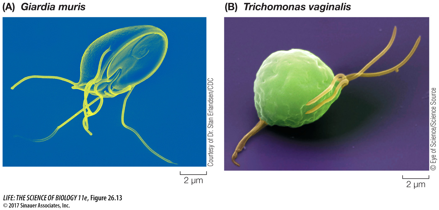
In addition to flagella and a cytoskeleton, the parabasalids have undulating membranes that also contribute to the cell’s locomotion. Trichomonas vaginalis (Figure 26.13B) is a parabasalid responsible for a sexually transmitted disease in humans. Infection of the male urethra, where it may occur without symptoms, is less common than infection of the vagina.
HETEROLOBOSEANS The amoeboid body form appears in several protist groups that are only distantly related to one another. The body forms of heteroloboseans, for example, resemble those of loboseans, an amoebozoan group that is not at all closely related to heteroloboseans (see the next section). Amoebas of the free-
EUGLENIDS AND KINETOPLASTIDS The euglenids and kinetoplastids together constitute a clade of unicellular excavates with flagella. Their mitochondria contain distinctive disc-
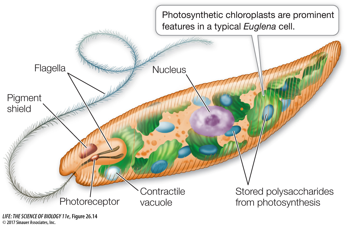
The flagella of euglenids arise from a pocket at the anterior end of the cell. Spiraling strips of proteins under the cell membrane control the cell’s shape. Some euglenids are photosynthetic. Figure 26.14 depicts a typical cell of the genus Euglena. Like most other euglenids, this common freshwater organism has a complex cell structure. It propels itself through the water with the longer of its two flagella, which may also serve as an anchor to hold the organism in place. The second flagellum is often rudimentary.
Media Clip 26.4 Euglenids
The euglenids have diverse nutritional requirements. Many species are always heterotrophic. Other species, including species of Euglena, are fully autotrophic in sunlight, using chloroplasts to synthesize organic compounds through photosynthesis. When kept in the dark, these euglenids lose their photosynthetic pigment and begin to feed exclusively on dissolved organic material in the water around them. A “bleached” Euglena resynthesizes its photosynthetic pigment when it is returned to the light and becomes autotrophic again. But Euglena cells treated with certain antibiotics or mutagens lose their photosynthetic pigment completely; neither they nor their descendants are ever autotrophs again. However, those descendants function well as heterotrophs.
The kinetoplastids are unicellular parasites with two flagella and a single, large mitochondrion. The mitochondrion contains a kinetoplast, a unique structure housing multiple circular DNA molecules and associated proteins. Some of these DNA molecules encode “guide proteins” that edit mRNA within the mitochondrion.
The kinetoplastids include several medically important species of pathogenic trypanosomes (Table 26.1). Some of these organisms are able to change their cell surface recognition molecules frequently, allowing them to evade our best attempts to kill them and thus eradicate the diseases they cause.
| Trypanosoma brucei | Trypanosoma cruzi | Leishmania major | |
|---|---|---|---|
| Human disease | Sleeping sickness | Chagas disease | Leishmaniasis |
| Insect vector | Tsetse fly | Assassin bugs (many species) | Sand fly |
| Vaccine or effective cure | None | None | None |
| Strategy for survival | Changes surface recognition molecules frequently | Causes changes in surface recognition molecules on host cell | Reduces effectiveness of macrophage hosts |
| Site in human body | Bloodstream; in final stages, attacks nerve tissue | Enters cells, especially muscle cells | Enters cells, primarily macrophages |
| Approximate number of deaths per year | 7,000 | 11,000 | 63,000 |