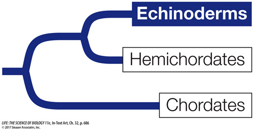
Echinoderms have unique structural features
In addition to pentaradial symmetry, adult echinoderms have two unique structural features. One is a system of calcified internal plates covered by thin layers of skin and some muscles. The calcified plates of most echinoderms are thick, and they fuse inside the entire body, forming an internal skeleton. The other unique feature of this group is a water vascular system, a network of water-
Members of one major extant echinoderm clade, the crinoids (sea lilies and feather stars), were more abundant and species-
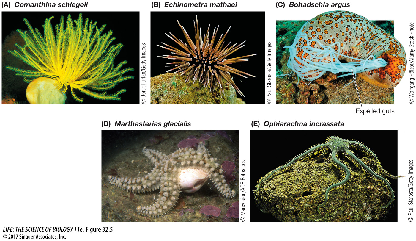
Although some crinoids are sessile, most surviving echinoderms are motile. The two main groups of motile echinoderms are the echinozoans (sea urchins and sea cucumbers) and the asterozoans (sea stars and brittle stars). Sea urchins are hemispherical in shape and lack arms (Figure 32.5B). They are covered with spines that are attached to the underlying skeleton with ball-
Sea cucumbers also lack arms, and their bodies are oriented in an atypical manner for an echinoderm (Figure 32.5C). The mouth is anterior and the anus is posterior (front and rear), in contrast to the oral–
Sea stars, popularly called starfish, are the most familiar echinoderms (Figure 32.5D). Their gonads and digestive organs are located in the arms, as seen in Figure 32.3B. Their tube feet serve as organs of locomotion, gas exchange, and attachment. Each tube foot of a sea star consists of an internal ampulla connected by a muscular tube to an external suction cup that can stick to the substrate. The tube foot is moved by expansion and contraction of the circular and longitudinal muscles of the tube. Brittle stars are similar in structure to sea stars, but their flexible arms are composed of jointed, hard plates (Figure 32.5E), their gonads and viscera are contained in the central disk, and their tube feet lack suction cups or ampullae.
Echinoderms use their tube feet in a great variety of ways to capture prey. Sea lilies, for example, feed by orienting their arms in passing water currents. Food particles then strike and stick to the tube feet, which are covered with mucus-
Most sea urchins capture phytoplankton with their tube feet or scrape algae from rocks with a complex rasping structure. Sea cucumbers capture food with their anterior tube feet, which are modified into large, feathery, sticky tentacles that can be protruded from the mouth. Periodically a sea cucumber withdraws the tentacles, wipes off the material that has adhered to them, and digests it.
Many sea stars use their tube feet to capture large prey such as polychaetes, gastropod and bivalve mollusks, small crustaceans such as crabs, and fishes. With hundreds of tube feet acting simultaneously, a sea star can grasp a bivalve in its arms, anchor the arms with its tube feet, and by steady contraction of the muscles in its arms, gradually exhaust the muscles the bivalve uses to keep its shell closed (see Figure 32.5D). To feed on the bivalve, the sea star can push its stomach out through its mouth and then through the narrow space between the two halves of the bivalve’s shell. The sea star’s stomach then secretes enzymes that digest the prey.
Media Clip 32.1 Sea Star Hunting Bivalves
Most of the 2,000 species of brittle stars ingest particles from the upper layers of sediments and assimilate the organic material from them, although some species filter suspended food particles from the water, and others capture small animals.