Body segmentation is an early feature of vertebrate development
The vertebrate body plan, like that of arthropods, consists of repeating segments that are modified during development. These segments are most evident as the repeating patterns of vertebrae, ribs, nerves, and muscles along the anterior–
As the neural tube forms, mesodermal tissues gather along the sides of the notochord to form separate, segmented blocks of cells called somites (Figure 43.13). Somites produce cells that will become the vertebrae, ribs, muscles of the trunk, and limbs.
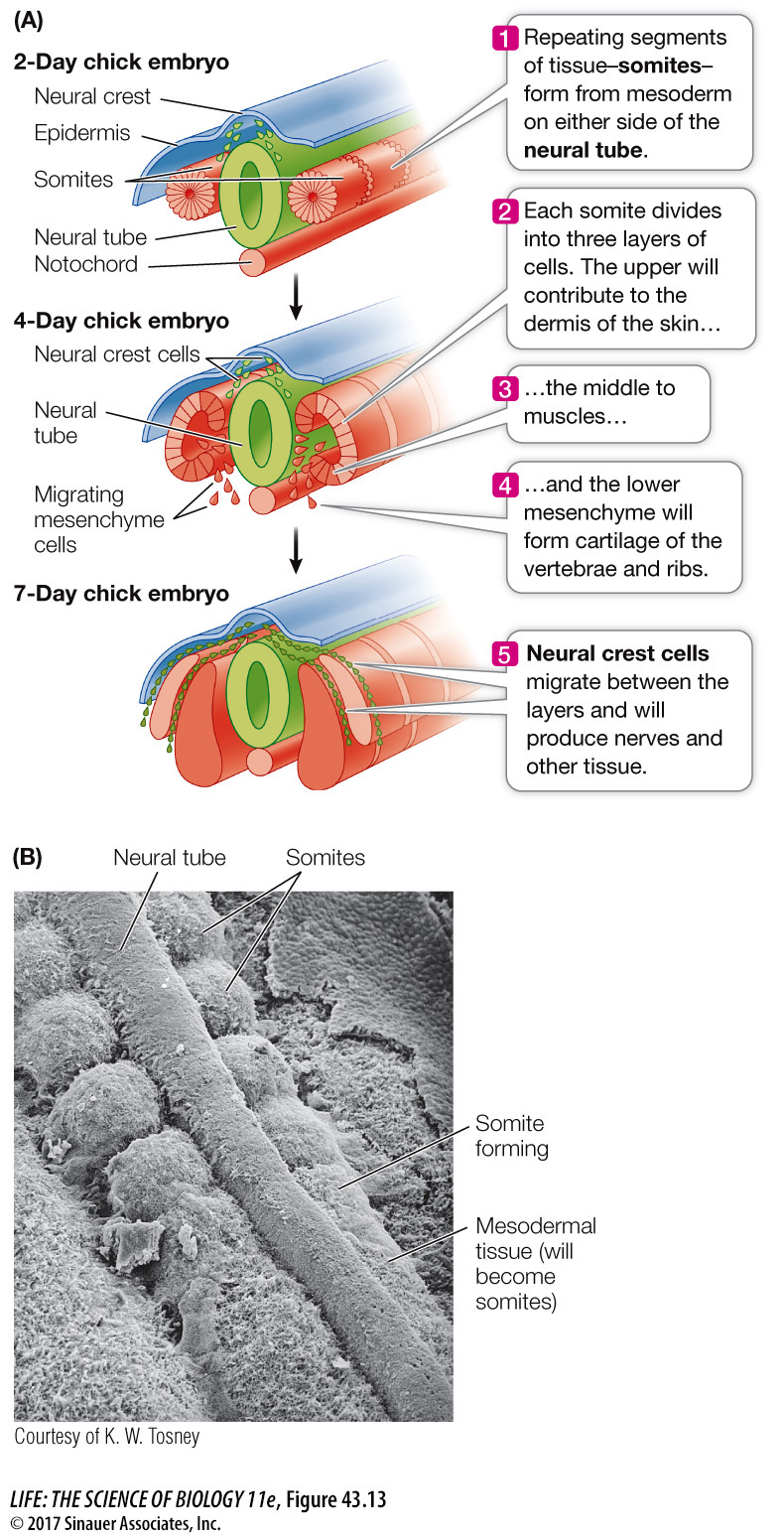
Nerves that connect the brain and spinal cord with tissues and organs throughout the body are also arranged segmentally. The somites help guide the organization of these peripheral nerves, but the nerves are not of mesodermal origin. As you saw above, when the neural tube fuses, the neural crest cells break loose and migrate inward between the epidermis and the somites and through the somites. These neural crest cells have diverse fates, including the development of peripheral nerves.
As development progresses, the different segments of the body change. Regions of the spinal cord differ, regions of the vertebral column differ in that some vertebrae grow ribs of various sizes and others do not, forelegs arise in the anterior part of the embryo, and hind legs arise in the posterior region.