Birds develop four extraembryonic membranes
The chicken provides a good example of how extraembryonic membranes form from the germ layers created during gastrulation. In the chick, four membranes form—the yolk sac, the allantoic membrane, the amnion, and the chorion. The yolk sac is the first to form, and it does so by extension of the hypoblast layer along with some adjacent mesoderm. The yolk sac grows to enclose the entire body of yolk in the egg (Figure 43.15). It constricts at the top to create a tube that is continuous with the gut of the embryo. However, yolk does not pass through this tube. Yolk is digested by the cells of the yolk sac, and the nutrients are transported to the embryo through blood vessels that form from mesoderm and line the outer surface of the yolk sac. The allantoic membrane is also an outgrowth of the extraembryonic endoderm plus adjacent mesoderm. It forms the allantois, a sac for storage of metabolic wastes.
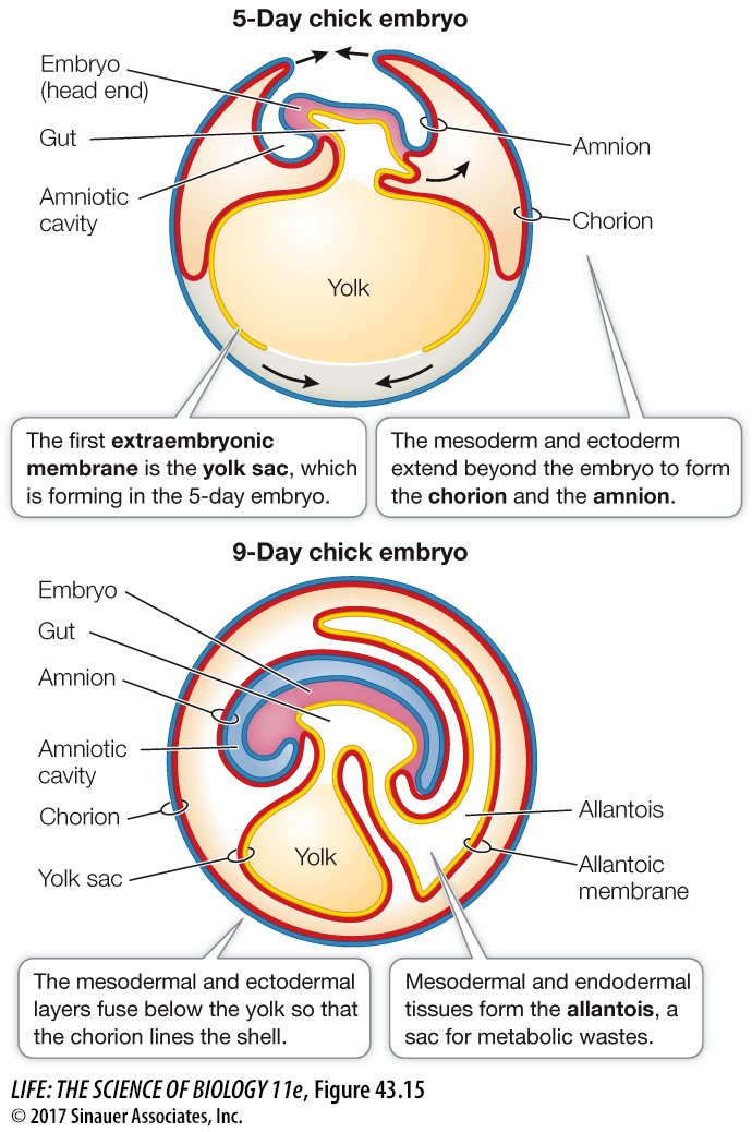
Figure 43.15 The Extraembryonic Membranes In birds (and other reptiles) and mammals, the embryo constructs four extraembryonic membranes. In birds, the yolk sac encloses the yolk, and the amnion and chorion enclose the embryo. Fluids secreted by the amnion fill the amniotic cavity, providing an aqueous environment for the embryo. The chorion, along with the allantoic membrane, mediates gas exchange between the embryo and its environment. The allantois stores the embryo’s waste products. (See also Figure 32.19.)
Activity 43.1 Extraembryonic Membranes
Page 933
Ectoderm and mesoderm combine and extend beyond the limits of the embryo to form the other extraembryonic membranes. Two layers of cells extend all along the inside of the eggshell, both over the embryo and below the yolk sac. Where they meet, they fuse, forming two membranes, the inner amnion and the outer chorion. The amnion surrounds the embryo, forming the amniotic cavity. The amnion secretes fluid into the cavity, providing a protective environment for the embryo. The outer membrane, the chorion, forms a continuous membrane just under the eggshell (see Figure 43.16) that limits water loss from the egg. The exchange of O2 and CO2 between the embryo and the environment requires diffusion of those gases across the chorion and the allantois.
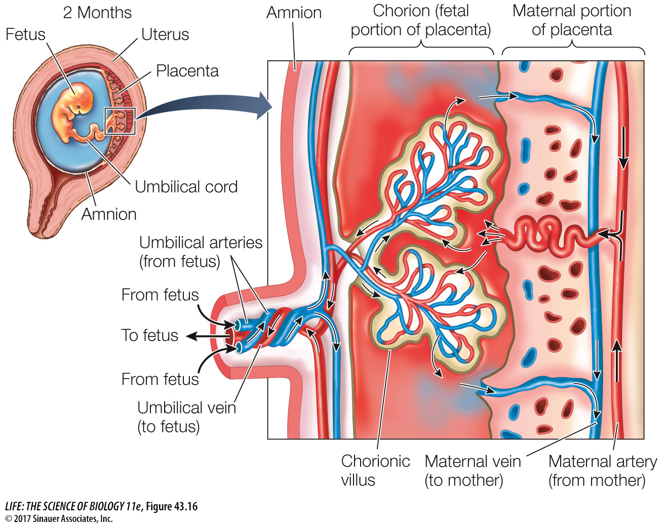
Figure 43.16 The Mammalian Placenta In humans and most other mammals, nutrients and wastes are exchanged between maternal and fetal blood in the placenta, which forms from the chorion and tissues of the uterine wall. The embryo is attached to the placenta by the umbilical cord. Embryonic blood vessels invade the placental tissue to form fingerlike chorionic villi. Maternal blood flows into the spaces surrounding the villi, and placental blood flows through the villi so nutrients and respiratory gases can be exchanged between the maternal and fetal blood.