Reflexes are controlled by simple circuits involving sensory neurons, interneurons, and effectors
A reflex is a behavioral or physiological response that does not require conscious information processing—it is autonomic, meaning an involuntary or unconscious reaction. Examples are salivating at the sight or smell of food, and the jerk of your lower leg when a physician taps your knee with a small rubber hammer. The knee-jerk reflex involves a very simple neural circuit involving just a few neurons that connect with each other in the spinal cord. Thus it is called a spinal reflex. A cross section of the human spinal cord reveals a central area of gray matter in the shape of a butterfly, surrounded by areas of white matter (see Figure 44.14). In the nervous system, gray matter is rich in neuronal cell bodies, and white matter contains myelinated axons. The gray matter of the spinal cord contains the cell bodies of the spinal neurons; the white matter contains myelinated axons that conduct information up and down the spinal cord.
Spinal nerves extend from the spinal cord at regular intervals on each side. Each spinal nerve has two roots, one connecting with the dorsal horn of the gray matter, the other with the ventral horn. The afferent (sensory) axons in a spinal nerve enter the spinal cord through the dorsal root, and the efferent (motor) axons leave through the ventral root. The neural circuit of the knee-jerk reflex is diagrammed in Figure 44.14. The tap of the physician’s rubber hammer stretches the tendon going over the knee. That tendon attaches the muscle of the upper leg to bone in the lower leg. Stretching the tendon stretches muscle fibers in the upper leg, and stretch receptors in that muscle transduce the physical stimulus into APs. The APs are then conducted by a sensory neuron into the dorsal horn of the spinal cord. That sensory neuron goes all the way to the ventral horn and synapses onto a motor neuron, which fires APs. The axon of that motor neuron travels out through the ventral horn of the spinal cord and extends all the way to the same muscle that initially was stretched, causing that muscle to contract.
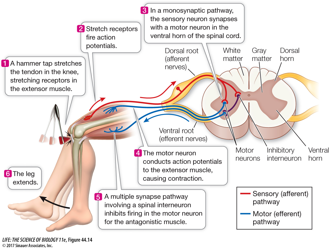
Figure 44.14 A Neural Network in the Spinal Cord Generates the Knee-Jerk Reflex Sensory (afferent) information enters the spinal cord through the dorsal horns, and motor (efferent) output leaves it via the ventral horns. Information travels to the brain in white matter tracts. Interneurons make connections within the spinal cord that result in a complex, coordinated behavior pattern.
Animation 44.4 Information Processing in the Spinal Cord
What is the function of the spinal knee-jerk reflex? This simple circuit senses increased load on a muscle and adjusts its level of contraction to match the load. Most spinal reflexes are more complex. For example, limb movement is controlled by antagonistic sets of muscles that work against each other. When one member of an antagonistic set of muscles contracts, it bends (flexes) the limb; it is therefore called a flexor. The antagonist muscle, the extensor, straightens (extends) the limb. For a limb to move, one muscle of the pair must relax while the other contracts. Thus sensory input that activates the motor neuron of one muscle also inhibits its antagonist. This coordination is achieved by an interneuron between the sensory neuron and the motor neuron of the antagonist muscle (see Figure 44.15). Thus the reciprocal inhibition of antagonistic muscles involves at least two synapses.
The withdrawal reflex is an example of a polysynaptic spinal reflex that involves many interneurons. When you step on a tack, you immediately pull back your foot, but you don’t fall over because many other muscles change your posture. The tack stimulates pain receptors in the foot, and the sensory neurons transmit APs into the dorsal horn of the spinal cord on the same side of the body. In the dorsal horn, these neurons synapse with a variety of interneurons. Some send the pain information through their axons to the brain, resulting in the conscious sensation of pain. But even before the brain is aware of the pain, other interneurons coordinate the withdrawal of the foot with actions of other muscles to shift your weight onto the other leg. Thus a rather complex suite of movements is coordinated by a network of interneurons in the spinal cord. By extension, you can appreciate how much more complex the neural networks are that enable you to execute complex movements in time with music and coordinated with another person—in other words, to dance.