Apply What You’ve Learned
Review
44.2
When gated ion channels are activated by electrical, chemical, or mechanical stimuli, permeability of the membrane to the respective ions changes, resulting in a change in membrane potential.
44.2
Rapid activation and inactivation of voltage-
44.3
Agonists and antagonists are drugs that target specific receptors.
Original Paper: Adamantidis, A. R., F. Zhang, A. M. Aravanis, K. Deisseroth and L. de Lecea. 2007. Neural substrates of awakening probed with optogenetic control of hypocretin neurons. Nature 450: 420−424.
A common question in neurobiology is whether or not neurons in a particular brain region control a specific physiological or behavioral response in the animal. Recording neural activity with electrodes may show a correlation between activity of the neurons and the response, but that does not prove that those neurons control the response. The best evidence for whether specific neurons control a specific response is obtained by stimulating the activity of the neurons and observing—
Dr. Karl Deisseroth’s lab at Stanford University developed a means of activating specific neurons with light. They called their method “optogenetics.” Algae have light-
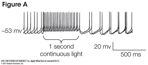
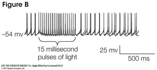
Using electrodes, the scientists recorded the action potentials of the hypocretin neurons when they were stimulated with light. Figure A shows the response of a neuron to 1 second of light, and Figure B shows the response to 15-
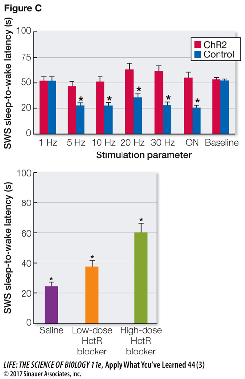
The researchers recorded sleep and wakefulness in the mice, and they stimulated the hypocretin neurons when the mice were asleep. The control mice received the same viral infection treatment, but the virus did not include the ChR2 gene. The time between the stimulation and the animal awakening (sleep-
Questions
1.
Describe the effect of 1 second of light on the membrane potential and the firing rate of the hypocretin neurons. What do these results suggest about the nature of the ChR2 protein and its response to light?
The 1-
2.
What is the evidence that light stimulation has a direct effect on the activity of the hypocretin neurons that are expressing ChR2?
The fact that the firing of the neurons followed precisely the trains of very short bursts of light (15 ms) indicates that the APs of the neurons were direct responses to the light.
3.
Describe the effect of light stimulation on the behavior of the mice. Did the frequency of stimulation matter?
The light stimulation of hypocretinergic neurons expressing ChR2 in sleeping mice shortened the latency to awakening. The same light stimulation had no effect on the mice not expressing ChR2. Above a frequency of 1 flash per second, the awakening response was not altered by the frequency of stimulation.
4.
What is the evidence that the effects of the stimulation were due to release of hypocretin by the stimulated neurons?
The fact that a blocker of the hypocretin receptor eliminated the awakening effect of the light stimulation supports the conclusion that the effect was mediated by the release of hypocretin by the stimulated neurons.
Go to LaunchPad for the eBook, LearningCurve, animations, activities, flashcards, and additional resources and assignments.