The structure of neurons reflects their functions
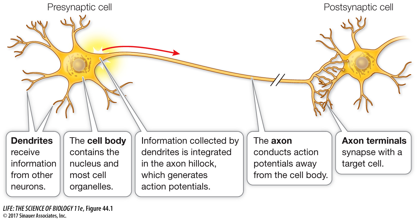
Neurons vary enormously in structure and appearance, but they have a basic structure that includes four regions (Figure 44.1).
A cell body contains the nucleus and most of the cell’s organelles.
Shrublike projections called dendrites (Greek dendron, “tree”) may extend from the cell body. Dendrites receive information from other neurons or sensory cells and bring it to the cell body.
In most neurons, one projection—
the axon—is much longer than the others. Axons carry information from the cell body to target cells. At the target cell, the axon divides into a spray of fine nerve endings. The tips of these tiny nerve endings have swellings called axon terminals.
Neurons have a wide variety of forms that reflect different functionalities (Figure 44.2). Neurons with few dendrites receive information from specific and limited sources, whereas neurons with large arrays of dendrites can collect and integrate information from a wide range of sources. Some neurons communicate over very short distances, but others communicate over extremely long distances and therefore have very long axons. For example, axons from neurons in your spinal cord control the muscles in your toes.
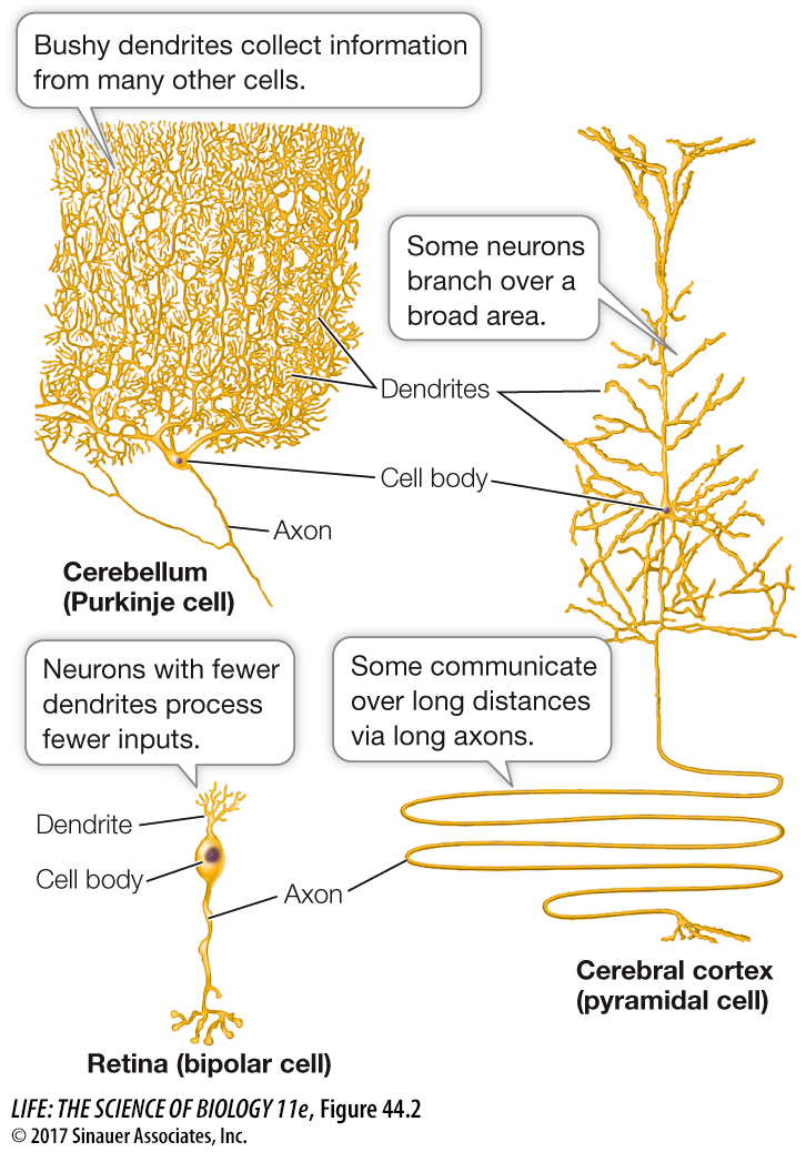
Q: Which of these neurons is likely to receive a greater number of inputs?
The Purkinje cell has the most dendrites and therefore is likely to receive more inputs than the pyramidal cell, and certainly more than the retinal cell.
Regardless of form and function, all neurons process and communicate information through changes in the electric potential across their membranes. As in all cells, the electric charge on the inside of the cell membrane of a neuron is slightly negative in comparison with the outside. The electric potential across the cell membranes of neurons can change in response to specific stimuli, and these electric potential changes can travel along the cell membrane. In most neurons, small changes in membrane electric potential generate large, rapidly reversed changes in membrane potential that are called action potentials (APs). Axons conduct APs over long distances.
In summary, a neuron receives specific stimuli (e.g., light, sound, pressure, chemicals) that cause changes in the electric potentials across the cell membranes of its dendrites. These electric signals from dendrites spread to and are averaged by the cell body. Changes in the electric potential of the membrane of the cell body at the base of its axon can cause an AP that is conducted along the axon to its terminals. APs can travel at speeds up to 100 meters per second (360 kilometers per hour), making it possible for an individual to sense, process, and act on information very quickly.
What happens when the AP reaches the axon terminals? Axon terminals come extremely close to the membrane of the target cell, which can be another neuron, a muscle cell, or a secretory cell. Structures called synapses form at these locations. The synapse transfers the information conveyed by the AP from the presynaptic cell to the postsynaptic cell (see Figure 44.1).
Synapses can be either electrical or chemical. Electrical synapses allow the AP to pass directly between two neurons. In vertebrates most synapses are chemical. At chemical synapses a space about 25 nanometers wide (about 1/2000th the width of a human hair) separates the presynaptic and postsynaptic membranes. An AP arriving at an axon terminal causes it to release chemical messenger molecules called neurotransmitters. Neurotransmitters diffuse across the synaptic space and bind to receptors on the cell membrane of the postsynaptic (target) cell. The binding of the neurotransmitter to its receptor alters the activity of the postsynaptic neuron. Some neurotransmitter–