Glia are the “silent partners” of neurons
The human brain has at least as many glial cells as neurons. The ratio differs in different brain areas, and in some areas is considerably greater than 1 to 1. A neurobiologist once said that “flashy neurons get all of the attention, but glial cells do most of the brain’s work and are the cause of many of its diseases.” It has been easier to study the functions of neurons because their APs can be observed. The mostly silent glia are more difficult to study, and therefore we know much less about them. Our knowledge of glia will grow enormously in the years to come.
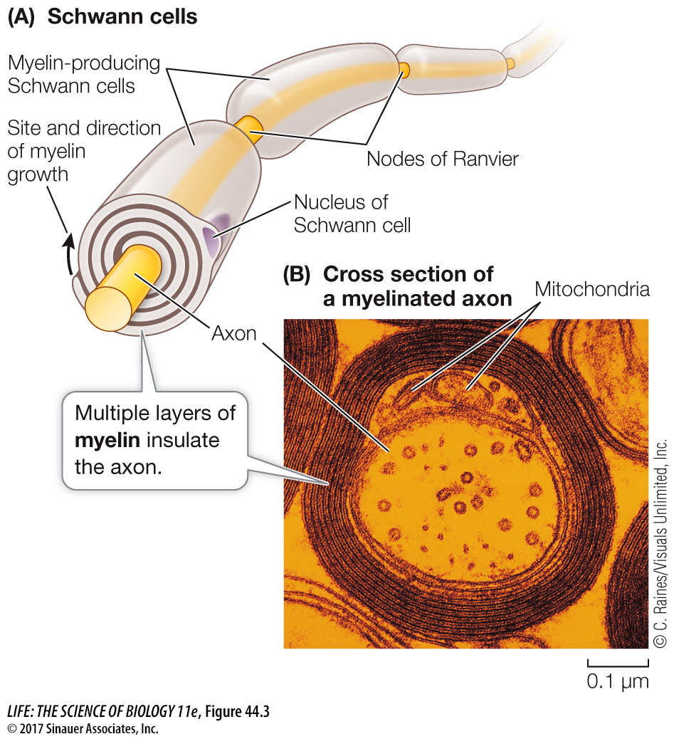
Like neurons, glia come in several forms and have diverse functions. As mentioned above, microglia are macrophages (see Figure 41.3) and provide immune defense responses for the nervous system. Macroglia include Schwann cells, oligodendrocytes, and astrocytes. In the brain and spinal cord, oligodendrocytes wrap around the axons of neurons, covering them with concentric layers of cell membrane. You can think of this wrapping as being insulation, like the insulation on electrical wires. It prevents electric current from leaking out of the axon. Outside the brain and spinal cord, Schwann cells provide this same function for the peripheral nerves that communicate between the brain and spinal cord and all parts of the body (Figure 44.3). Myelin is the wrapping produced by oligodendrocytes and Schwann cells, and it gives many parts of the nervous system a glistening white appearance. Not all axons are myelinated, but those that are conduct APs more rapidly than can axons that are not myelinated, for reasons described in Key Concept 44.2.
Diseases that affect myelin can be devastating because they impair conduction of APs. The most common of these demyelinating diseases is multiple sclerosis (MS)—literally “multiple scars”—which occurs in about 1 in 700 people in the United States. The cause of MS is not known, but it involves inflammatory autoimmune damage to the myelin in the brain and spinal cord. The symptoms of the disease depend on where in the nervous system the myelin is damaged. Motor impairment is common. An example of a demyelinating disease that attacks myelin outside the brain and spinal cord is Guillain–
The third type of macroglia, astrocytes (so named because they look like stars), contribute to the blood–
In addition to their role in the blood–
They can take up neurotransmitter that has been released into the synapse and thereby control communication between the pre-
and postsynaptic cells. They can supply neurons with nutrients. Neurons have no energy reserves, but astrocytes store glycogen that they can break down to supply the neurons with fuel.
They have signaling properties. Even though most astrocytes do not generate APs, they do release neurotransmitters that can alter the activities of neurons.
They aid in the repair and regeneration of neurons.
They make contact with both blood vessels and neurons and can therefore signal changes in the composition of the blood.
Astrocytes play crucial yet poorly understood roles in modulating synapse activity. The projections of a single astrocyte may make contact with more than 100,000 synapses. The contact of the astrocyte with the neuronal components of the synapse is so intimate that it has inspired the concept of the tripartite synapse—the idea that a synapse includes not only the pre-
Although astrocytes do not generate APs, they do communicate with each other. They are connected to each other through electrical synapses, and the electric signals traveling across these synapses cause changes in the Ca2+ content of the postsynaptic astrocyte. When Ca2+-sensitive dyes are applied to neural tissue, Ca2+ waves can be seen traveling through extensive networks of astrocytes. The functions of these Ca2+ waves are not understood.