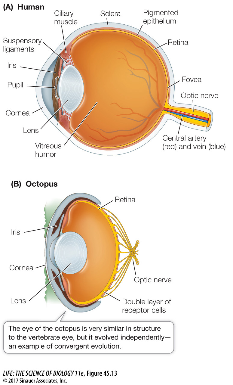
Image-forming eyes evolved independently in vertebrates and cephalopods
Both vertebrates and cephalopod mollusks (such as squid and octopus) have eyes with exceptional abilities to form detailed images of the visual world. Like cameras, both of these eye types focus inverted images on an internal surface that is sensitive to light. Considering that they evolved completely independently of each other, their degree of similarity is remarkable (Figure 45.13).
Figure 45.13Convergent Evolution of Eyes The lenses of vertebrate (A) and cephalopod (B) eyes focus images on layers of photoreceptor cells.
Q: Humans commonly experience “floaters” in their eyes—tissue fragments that float around in the field of vision. Where in the structure of the eye would you expect floaters to exist?
Floaters in the eye would have to be in the vitreous humor.
The vertebrate eye (see Figure 45.13A) is a spherical, fluid-filled structure bounded by a tough connective tissue layer called the sclera. The sclera at the front of the eye forms the transparent cornea, through which light passes to enter the eye. Just inside the cornea is the pigmented iris, which gives the eye its color. The iris controls the amount of light that reaches the photoreceptor cells at the back of the eye, just as the diaphragm of a camera controls the exposure. The central opening of the iris is the pupil. The iris is under neural control. In bright light, the iris constricts and the pupil is very small. As light levels fall, the iris opens and the pupil enlarges.
Behind the iris is the crystalline protein lens that makes fine adjustments in the focus of images falling on the photosensitive layer—the retina—at the back of the eye. The cornea and the gel-like mass (vitreous humor) within the eye bend light rays passing through them so that they are focused on the retina. The lens makes fine adjustments to the focus and allows the eye to accommodate—that is, to focus on objects at various locations in the near visual field. To focus a camera on objects close at hand, you adjust the distance between the lens and the internal surface sensitive to light. Fish, amphibians, and non-avian reptiles accommodate in a similar manner, moving the lenses of their eyes closer to or farther from their retinas. Mammals and birds use a different method; they alter the shape of the lens.
Page 974
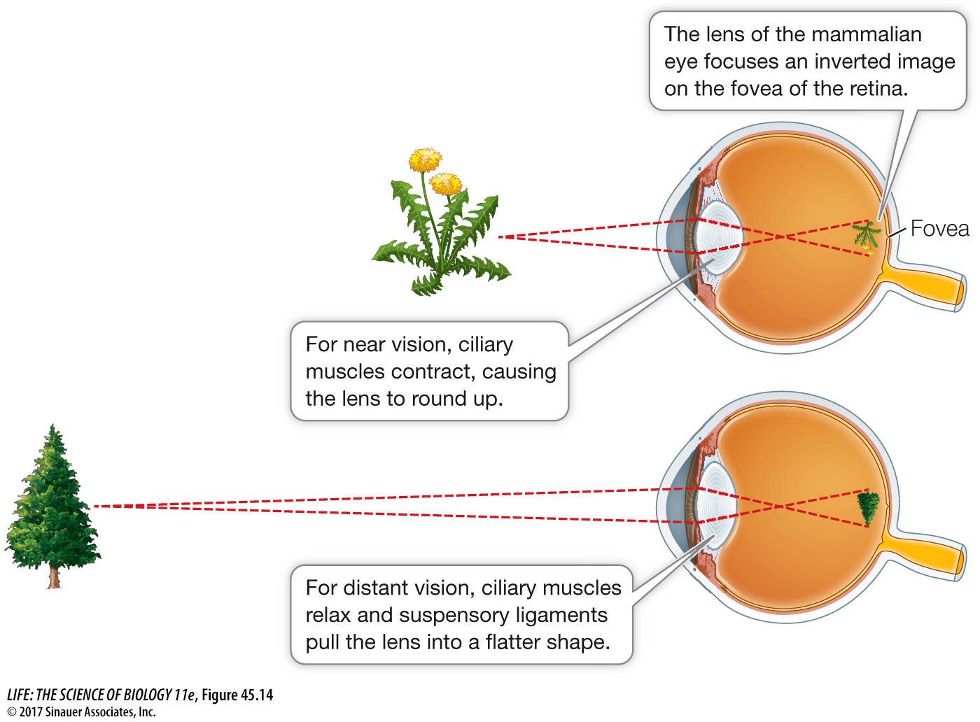
The mammalian lens is contained in a connective tissue sheath that tends to keep it in a spherical shape, but the sheath is attached to suspensory ligaments that pull the lens into a flatter shape. Circular ciliary muscles counteract the pull of the suspensory ligaments, permitting the lens to round up. When the ciliary muscles are at rest, the flatter lens has the correct optical properties to focus distant images on the retina. Contracting the ciliary muscles rounds up the lens, changing its light-bending properties to bring close images into focus (Figure 45.14).
Figure 45.14Staying in Focus Mammals and birds focus their eyes by changing the shape of the lens depending on the eye’s distance from the object of focus.
Lenses become less elastic with age, so we lose the ability to focus on objects close at hand without the help of corrective lenses. Most people over the age of 45 need the assistance of reading glasses or bifocal lenses.