The visual system is an example of information integration by the cerebral cortex
The visual system is one of the most-
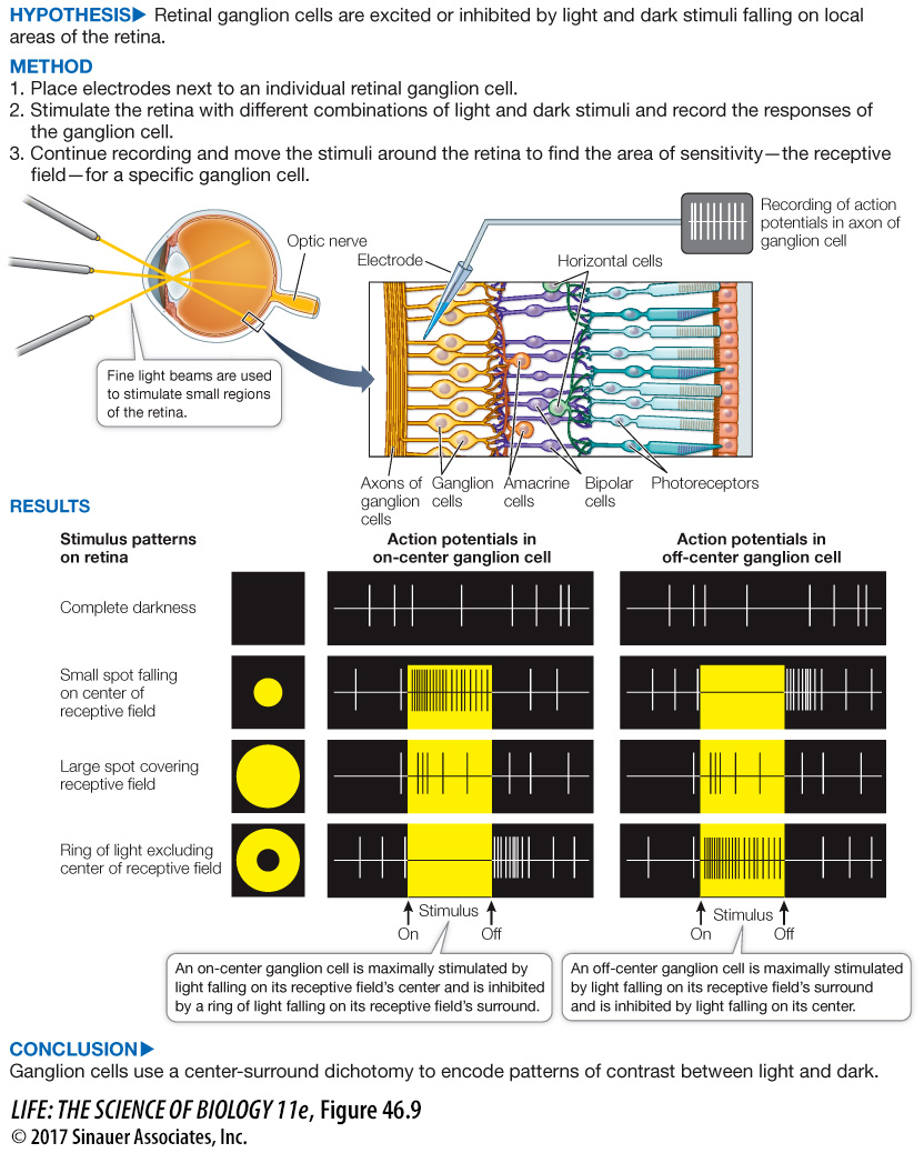
RETINAL RECEPTIVE FIELDS Key Concept 45.4 described how a retinal ganglion cell collects information from several photoreceptors—
The question of what information the retinal ganglion cell extracts from the photoreceptors was addressed in classic experiments by Stephen Kuffler, then at Johns Hopkins University. He used electrodes to record the activity of single ganglion cells of cat eyes while stimulating their retinas with spots of light (Figure 46.9). Kuffler’s experiments revealed that each ganglion cell has a well-
The receptive fields of most ganglion cells are circular, but whether a spot of light falling on a receptive field excites or inhibits its ganglion cell depends both on the nature of the receptive field and on where the spot of light falls on it. Receptive fields have a center and a concentric surround, and can be either “on-
Center effects are always stronger than surround effects. Thus a small dot of light directly on the center of a receptive field has the maximum effect, and a larger light stimulus illuminating the center and parts of the surround has a smaller effect. A uniform patch of light falling equally on the center and surround has very little effect on the firing rate of the ganglion cell for that receptive field. The center and surround almost exactly cancel one another, even though the area of the surround is much larger.
Activity 46.2 Visual Receptive Fields Simulation
As is seen in Figure 46.9, photoreceptors synapse onto bipolar cells and bipolar cells onto ganglion cells. This pattern of connectivity describes the relationship between the photoreceptors in the center of a receptive field. The photoreceptors in the surround area modify communication between the center photoreceptors and their bipolar cells through the lateral connections of horizontal cells and amacrine cells. Thus the receptive field of a ganglion cell results from a pattern of synapses between photoreceptors, horizontal cells, amacrine cells, and bipolar cells. A general lesson to learn from this seemingly confusing chain of events is that inhibition can be as important as excitation in neural circuits.
experiment
Figure 46.9 What Does the Eye Tell the Brain?
Original Paper: Kuffler, S. W. 1953. Discharge patterns and functional organization of mammalian retina. Journal of Neurophysiology 16: 37–
Stephen Kuffler’s experiments recorded the activity of single ganglion cells in the eyes of cats. These groundbreaking experiments revealed the existence of a circular receptive field for each of the retina’s ganglion cells. Signals from photoreceptor cells in a receptive field are either excitatory or inhibitory to the ganglion cell, which sends action potentials via the optic nerve to the brain.
Q: Why is there a blind spot in each eye?
The blind spot in each eye corresponds to the location on the retina where the axons of the ganglion cells leave the retina to form the optic nerve. There are no photoreceptors at those locations.
In summary, the neural circuitry of the retina results in the generation of signals in the axons of the optic nerve that communicate simple information about the contrasting patterns of light and dark falling on different parts of the retina. But once the action potentials in the optic nerve reach their destinations, how does the brain integrate them to construct visual images of the outside world?
Animation 46.1 Information Processing in the Retina
RECEPTIVE FIELDS OF CELLS IN THE VISUAL CORTEX The axons of the optic nerves terminate in a region of the thalamus that is a relay station receiving information from both the right and left eyes. From the thalamus, the information encoded in the activity of axons in the optic nerves is relayed to the visual cortex in the occipital lobes at the back of the brain. In the 1960s David Hubel and Torsten Wiesel of Harvard University studied the activity of neurons in the visual cortex by shining spots and bars of light on retinas while recording the activities of single cells in the cortex. They found that many neurons in the visual cortex, like retinal ganglion cells, have receptive fields with regions that are mutually antagonistic.
Hubel and Weisel discovered that neurons in the visual cortex respond selectively to bars of light of different orientations falling on the retina, and in some cases to movement of those bars of light in different directions. The concept that emerges from these experiments is that the brain assembles a mental image of the visual world by analyzing edges in patterns of light falling on the retina. Each retina sends a million axons to the brain, but there are hundreds of millions of neurons in the visual cortex. The action potentials from one retinal ganglion cell are received via the thalamus by hundreds of cortical neurons, each responsive to a different combination of orientation, position, color, and movement of contrasting lines in the patterns of light and dark falling on the retina.