The anatomical organization of the CNS emerges during development
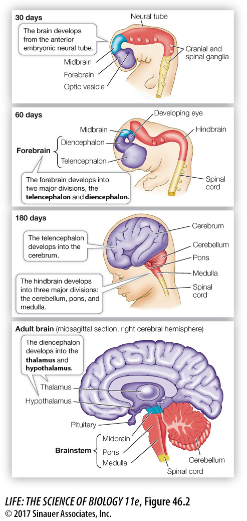
Early in the development of a vertebrate embryo, a tube of neural tissue forms (see Key Concept 43.4). At its anterior end, this neural tube forms three swellings that become the hindbrain, midbrain, and forebrain. The rest of the neural tube becomes the spinal cord. Peripheral nerves sprout from the midbrain and hindbrain (the cranial nerves) and from the spinal cord (the spinal nerves). From these early stages we see the linear axis of information flow in the nervous system. Although the developing brain will fold and become a complex structure, the information flow in the adult nervous system will follow paths that emerge from the simple, linear neural tube.
Each of these three regions of the embryonic brain develops into several structures in the adult brain (Figure 46.2). From the embryonic midbrain come structures that integrate information from the different senses and coordinate motor responses. From the hindbrain come the medulla, the pons, and the cerebellum. The medulla is continuous with the spinal cord, the pons is anterior to the medulla, and the cerebellum is a dorsal outgrowth of the pons. The medulla and pons contain distinct groups of neurons involved in controlling physiological functions such as breathing, circulation, and basic motor patterns such as swallowing and vomiting. All information traveling between the spinal cord and higher brain areas must pass through the pons, the medulla, and the midbrain, which are collectively known as the brainstem.
One function of the cerebellum is coordinating muscle activity and maintaining balance. It is like the director of a movie; the cerebellum receives a “script” of the commands going to the muscles from higher brain areas, and it receives information about the actual performance coming up the spinal cord from the “actors”—the joints and muscles. The cerebellum compares the “script” with the performance and refines motor commands accordingly. Damage to the cerebellum results in loss of fine motor control and coordination.
The embryonic forebrain develops a central region called the diencephalon and a surrounding structure called the telencephalon. The diencephalon is the core of the forebrain and consists of an upper structure, the thalamus, and a lower structure, the hypothalamus. The thalamus is the final relay station for sensory information going to the telencephalon. The hypothalamus receives a lot of physiological information of which we are not conscious, and it uses that information to regulate many physiological functions and biological drives. A major function of the hypothalamus is to control the pituitary gland (see Key Concept 40.2).
The embryonic telencephalon gives rise to the cerebrum, consisting of the left and right cerebral hemispheres. The outer layer of the cerebrum is the cerebral cortex, a thin layer rich in cell bodies. If we compare vertebrate groups from fish through amphibians, reptiles, and mammals, the cerebrum increases in size, complexity, and importance—