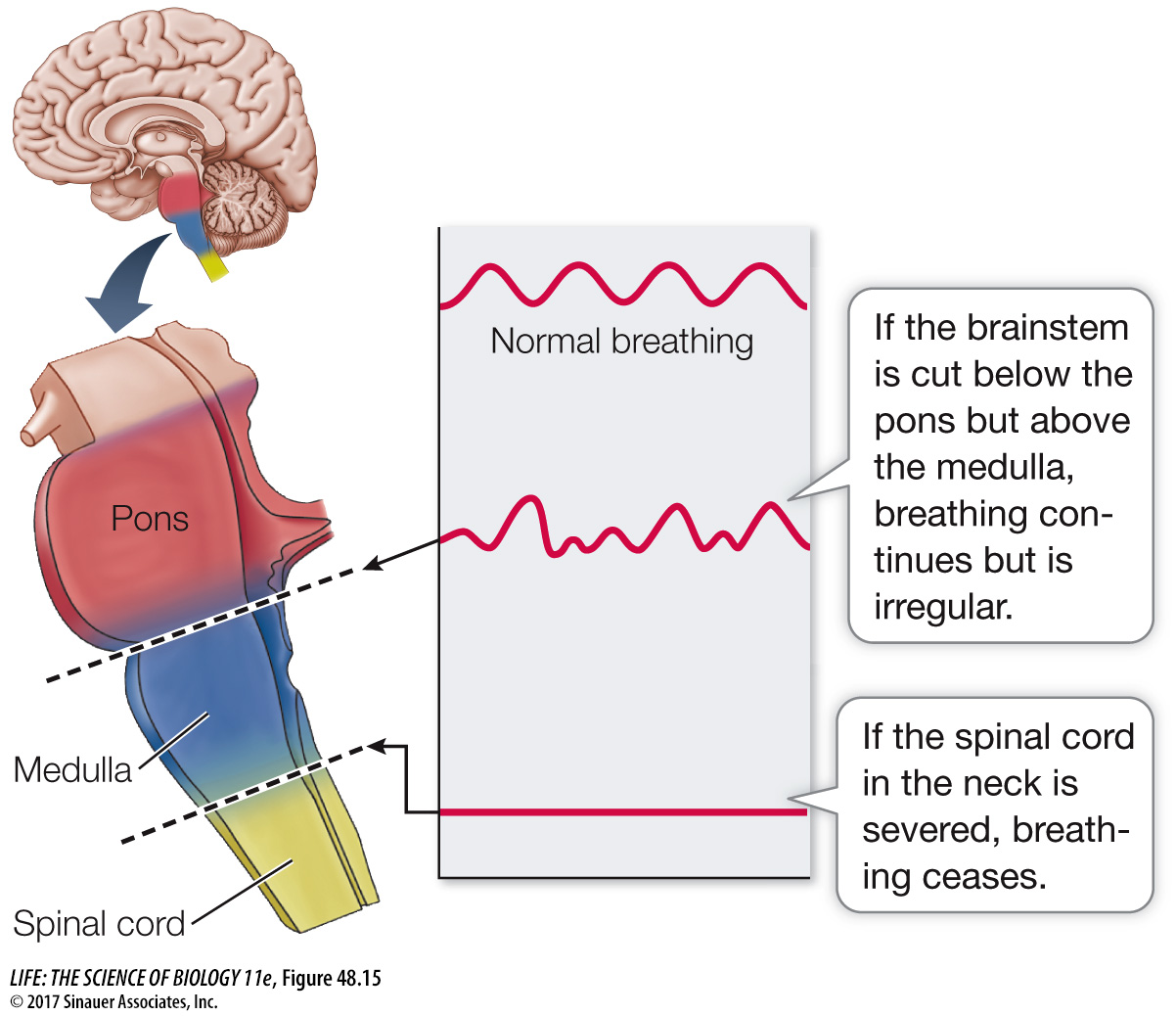
Breathing is driven by a rhythm-generating neural circuit in the brainstem, but where? Breathing ceases if the spinal cord is severed in the neck region, showing that breathing is generated in the brain. If the brainstem is cut just above the pons, breathing is normal. If the brainstem is cut between the pons and the medulla (the segment of the brainstem just above the spinal cord), the breathing pattern becomes irregular (Figure 48.15). Electrophysiological recordings reveal a group of respiratory motor neurons in the dorsal medulla that increase their firing rates just before an inhalation begins. The axons of these neurons leave the CNS in the neck region to form the phrenic nerve, which innervates the diaphragm. As more and more of these neurons fire—and fire faster and faster—the diaphragm contracts. All of a sudden the neurons stop firing, the diaphragm relaxes, and exhalation begins. Exhalation is usually a passive process that depends on the elastic recoil of the lung tissues. Another group of respiratory motor neurons is found in the ventral medulla that becomes active when breathing demand is high. These motor neurons communicate through thoracic spinal nerves to the intercostal muscles. By expanding and contracting the rib cage, the intercostal muscles increase both the inhalation and the exhalation volumes.
Neurons in the lower region of the pons help regularize the basic respiratory rhythm. Still higher brain areas modify breathing to accommodate speech, ingestion of food, coughing, and emotional states.