Electrical properties of ventricular muscles sustain heart contraction
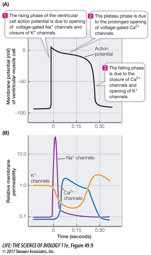
Electrical properties of ventricular muscle fibers allow them to contract for about 300 milliseconds—much longer than skeletal muscle fibers. As in neuronal and skeletal muscle action potentials, the rising phase of the ventricular muscle cell action potentials is due to the opening of voltage-gated Na+ channels. Unlike neurons and skeletal muscle fibers, however, ventricular muscle cells remain depolarized for a long time. This extended plateau of the action potential is due to sustained opening of voltage-gated Ca2+ channels (Figure 49.9). Like other muscle, cardiac muscle is stimulated to contract when Ca2+ is available to bind with troponin (see Figure 47.6). As long as Ca2+ remains in the sarcoplasm, the ventricular muscle cells continue to contract.
Figure 49.9The Action Potential of Ventricular Muscle Fibers(A) The three phases of the action potential of ventricular muscle fibers are due to the opening and closing of voltage-gated channels. (B) At the initiation of the action potential, voltage-gated Na+ channels open rapidly but briefly. At the same time, but more slowly, K+ channels are closing and Ca2+ channels are opening. The open Ca2+ channels sustain the depolarization. Repolarization occurs when the Ca2+ channels close, and the slow opening of the K+ channels also contributes to the repolarization.
Q: Why is the broadening of the ventricular muscle action potential important?
The breadth of the ventricular muscle action potential is important because it determines the duration of systole and therefore the time available for the emptying of the heart. More complete emptying means greater stroke volume.
To terminate systole and allow the ventricles to fill again, Ca2+ must be rapidly cleared from the sarcoplasm of the ventricular cells. Ca2+ pumps in the sarcoplasmic reticulum membrane and the cell membrane actively transport Ca2+ ions out of the sarcoplasm and into the sarcoplasmic reticulum or into the intersitial fluid. Thus Ca2+ in the sarcoplasm is maintained at a low level until the next action potential triggers another round of Ca2+ release and muscle contraction. The rate of cycling of Ca2+ into and out of the sarcoplasm puts limits on the heart rate and strength of contraction of the ventricle.