The vertebrate gut consists of concentric tissue layers
Tissues of the vertebrate gut are arranged in concentric layers that have a similar organization throughout its length (Figure 50.10). The mucosa lines the internal cavity, or lumen,of the gut. The mucosa consists of delicate epithelial cells with underlying connective tissue. Some cells of this mucosal epithelium secrete mucus to lubricate and protect the walls of the gut; some secrete digestive enzymes; and some secrete hormones. Mucosal epithelial cells in the stomach secrete hydrochloric acid, and as we noted in Key Concept 50.1, some secrete intrinsic factor to aid the absorption of vitamin B12. In some regions of the gut, nutrients are absorbed by mucosal epithelial cells. The apical cell membranes of these absorptive cells have microvilli that increase the surface area over which absorption can take place (see Figure 50.8C).
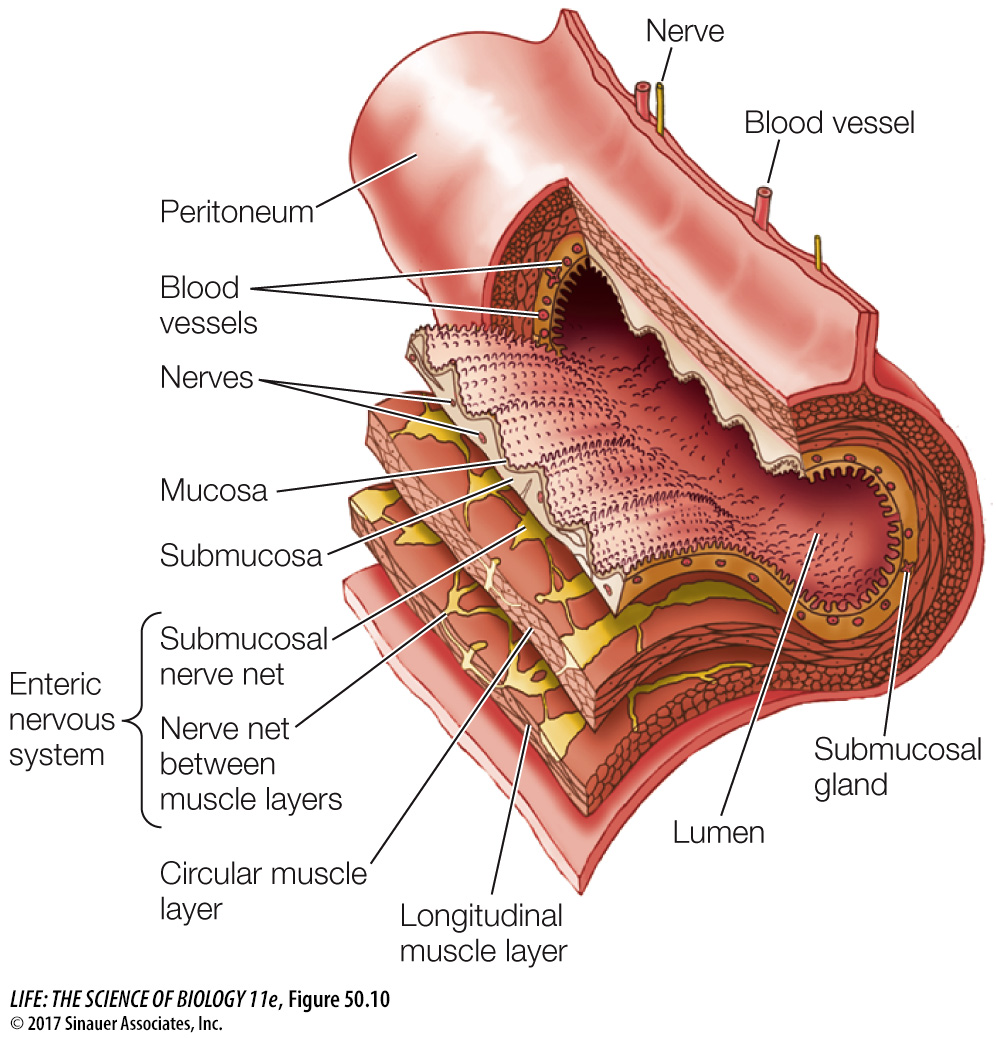
At the base of the mucosa there is a thin layer of smooth muscle cells with random orientations. The function of this cell layer is to move the mucosal epithelium to maximize its contact with the gut contents. Just under the mucosa is the submucosal tissue layer where blood and lymph vessels pick up nutrients to transport to the rest of the body. The submucosa also contains a network of nerves; the neurons in this network have sensory functions (responsible for stomach aches) and also control various secretory functions of the gut.
External to the submucosa are two layers of smooth muscle responsible for large movements of the gut. Innermost is the circular muscle layer, with its cells oriented around the gut. Outermost is the longitudinal muscle layer, with its cells oriented along the length of the gut (see Figure 50.10). The circular muscles constrict the gut, and the longitudinal muscles shorten it. Between the two layers of smooth muscle is another nerve network that controls and coordinates the movements of the gut. The coordinated activity of the two smooth muscle layers mixes contents of the gut and moves it continuously toward the anus.
Nerve nets in the submucosa and between the smooth muscle layers are called the enteric nervous system, and they are unusual. The cell bodies of most peripheral nerves are in the central nervous system (CNS)—in the brain or spinal cord. The only exceptions are the postganglionic cells of the autonomic nervous system and the cells of the enteric nervous system. In addition, most neurons of the peripheral nervous system either receive synapses from neurons in the CNS or contribute synapses to neurons in the CNS. In contrast, most of the neurons in the enteric nervous system form synapses only with other neurons in their network. Thus they are responsible for communication within the gut. The CNS can influence activity in the enteric nervous system and receive information from it, but the gut truly has “a mind of its own.”
A tissue membrane called the peritoneum surrounds the gut and most of the organs of the abdominal cavity as well as lining the wall of the cavity. The peritoneum includes connective and epithelial tissues that secrete a lubricating fluid that enables the organs to easily slide against each other in the body cavity.