The loop of Henle creates a concentration gradient in the renal medulla
Humans can produce urine that is four times more concentrated than their blood plasma. The vampire bat you encountered at the beginning of this chapter can produce urine that is 15 times more concentrated than its blood plasma (Investigating Life: How Can Vampire Bats Use Blood as Fast Food?). The concentrating ability of the mammalian kidney arises from a countercurrent multiplier mechanism made possible by the anatomical arrangement of the loops of Henle. The term “countercurrent” refers to the opposing directions of fluid flow in the descending and ascending limbs. The term “multiplier” refers to the ability of this system to create a solute concentration gradient in the renal medulla.
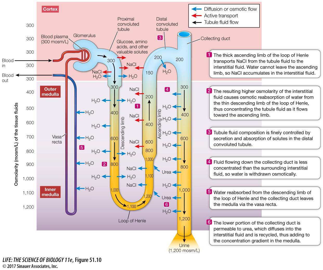
The loops of Henle do not themselves produce concentrated urine; rather, they increase the osmolarity of the extracellular fluid in the medulla in a graduated way. In humans, for example, the extracellular fluid at the top of the medulla bordering the cortex will be about 300 mosm/L (the concentration of blood plasma). But at the bottom of the medulla, where the loops of Henle make their hairpin turns, the extracellular fluid can be 1,200 mosm/L (Focus: Key Figure 51.10). How do the loops produce this effect?
focus: key figure
Q: What would be the effect of a drug called a “loop diuretic” that blocks the Na+ transport in the thick ascending limb of the loop of Henle?
Blocking the Na+ transport out of the thick ascending limb of the loop of Henle would result in a loss or a decrease (depending on the extent of the drug action) of the concentration gradient in the medulla. As a result, the tubular fluid in the collecting duct could not be concentrated, so urine flow would increase and there would be increased loss of water.
Animation 51.1 The Mammalian Kidney
The cells that make up the different segments of the loop of Henle differ anatomically and functionally. Cells of the descending limb and the initial cells of the ascending limb are thin, with no microvilli and few mitochondria. They are not specialized for transport. Partway up the ascending limb, the cells become specialized for active transport. These cells are thick and have many mitochondria. Accordingly, the segments of the loop of Henle are named the thin descending limb, the thin ascending limb, and the thick ascending limb (see Figure 51.9C).
The countercurrent multiplier mechanism is best understood by first considering events occurring in the thick ascending limb (see Figure 51.10, note 1). The cells of the thick ascending limb reabsorb Na+ and Cl– from the tubule fluid and move it into the interstitial fluid. (In the following discussion, we will distinguish between the two components of extracellular fluid—
The thin descending limb, in contrast, is highly permeable to water but not very permeable to Na+ and Cl–. Since the local interstitial fluid has been made more concentrated by the Na+ and Cl– reabsorbed from the neighboring thick ascending limb, water is withdrawn osmotically from the fluid in the descending limb. Therefore the fluid in the descending limb becomes more concentrated as it flows toward the hairpin turn at the bottom of the renal medulla (see Figure 51.10, note 2).
The thin ascending limb, like the thick ascending limb, is not permeable to water. It is, however, permeable to Na+ and Cl–. As the concentrated tubule fluid flows up the thin ascending limb, it is more concentrated than the surrounding interstitial fluid, so Na+ and Cl– diffuse out. When the tubule fluid reaches the thick ascending limb, active transport continues to move Na+ and Cl– from the tubule fluid to the interstitial fluid.
Because of this countercurrent multiplier mechanism, the tubule fluid reaching the distal convoluted tubule is less concentrated than the blood plasma (see Figure 51.10, note 3), and the solutes that have been left behind in the renal medulla have created a concentration gradient in the interstitial fluid of the medulla (indicated by the background color gradient in Figure 51.10).
You may wonder why the blood flow through the medulla does not wash out the concentration gradient established by the loops of Henle. The parallel arrangement of the descending and ascending peritubular capillaries in the medulla—