Blood osmolarity and blood pressure are regulated by ADH
Cells in the hypothalamus can stimulate the release of a hormone called antidiuretic hormone (ADH, also called vasopressin) from the posterior pituitary. ADH stimulates cells of the collecting duct to insert aquaporins (water channels) into their cell membranes. The aquaporins increase the permeability of these membranes to water, and therefore more water is reabsorbed from the collecting duct fluid into the interstitial fluid of the renal medulla. The higher the circulating levels of ADH, the greater the number of aquaporins. Various factors can stimulate or inhibit the release of ADH. Of key importance to kidney function are (1) osmoreceptors that monitor blood osmolarity and (2) stretch receptors that monitor blood pressure (Figure 51.14).
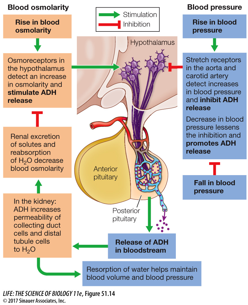
Osmoreceptor neurons in the hypothalamus are activated by a rise in blood osmolarity, and they increase the release of ADH. ADH helps regulate blood osmolarity by controlling water reabsorption. The osmoreceptors also stimulate thirst. The resulting water retention and water intake dilute the blood as they expand blood volume.
Stretch receptors in the walls of the aorta and the carotid arteries (see Figure 49.19) that detect an increase in blood pressure will inhibit the release of ADH. With less circulating ADH, less water is reabsorbed, which increases urine volume and decreases blood volume, hence lowering blood pressure.
If blood pressure falls, as when you lose blood volume through hemorrhage or excessive evaporative water loss, activity of the stretch receptors in the aorta and carotid arteries decreases. Input via cranial nerves to the hypothalamus from these receptors inhibits the release of ADH, so when the firing rates of these stretch receptors fall, ADH release increases. More ADH results in more efficient water reabsorption and therefore a protection of blood volume and blood pressure.
Alcohol inhibits ADH release, explaining why excessive beer drinking leads to excessive urination and dehydration, which contributes to the symptoms of a hangover.
As already mentioned, the presence of aquaporins in cell membranes determines their water permeability. Aquaporins play an important and unique role in the collecting duct. Several members of the aquaporin family of water channels are found in the cell membranes of the collecting duct cells. At least two aquaporins (AQP-
experiment
Figure 51.15 ADH Induces Insertion of Aquaporins into Cell Membranes
Original Paper: Nielsen, S., C. L. Chou, D. Marples, E. I. Christensen, B. K. Kishore and M. A. Knepper. 1995. Vasopressin increases water permeability of kidney collecting duct by inducing translocation of aquaporin-
Aquaporin proteins make some regions of renal tubules permeable to water. One aquaporin, AQP-
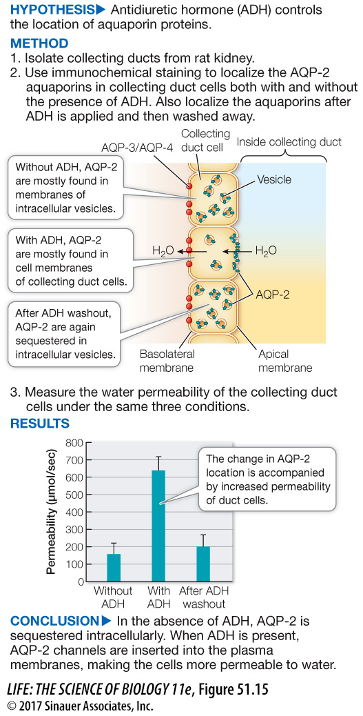
When ADH levels are low, such as when a person is well hydrated, most of the AQP-