Chapter 15
RECAP 15.1
Point mutations that cause phenotypic changes could have resulted in a different amino acid in the encoded protein that consequently changes a protein’s function; changed a promoter so a gene’s expression is significantly altered; or created a stop codon that terminates expression prematurely, resulting in a shorter nonfunctional protein. Point mutations that are phenotypically silent may arise in codons where redundancy ensures no amino acid change; cause codon changes that result in amino acid changes that are not significant to protein function; or occur in noncoding regions of DNA, such as introns.
Page A-16See Figure 15.4. Deletions are missing part of a chromosome; duplications have an extra copy (or copies) of a chromosomal region; in inversions, a chromosome region is out of sequence; and in translocations, a piece of one chromosome breaks off and attaches to another chromosome.
Spontaneous mutation occurs without an external agent causing it. Example: tautomeric shift of A, so that at DNA replication A base-
pairs not with T but with C. In an induced mutation, an environmental agent changes DNA. Example: nitrous acid deaminates C to U, so that at DNA replication, instead of C pairing with G, it is U pairing with A. C can be methylated to 5-
methylcytosine. When deaminated spontaneously or by a mutagen, this base forms T. This is a normal base and is not removed by DNA repair. Other base changes are repaired.
RECAP 15.2
The amino acid sequence produced by the mutant allele is Leu-
Ile- Ser- Ile- Ala. This is a missense mutation. The mutation replaces proline with serine. Proline is a nonpolar amino acid that is usually part of bends or loops in a protein; serine is a polar amino acid with a smaller side chain. The mutation is likely to affect enzyme activity because it is likely to affect protein structure.
Simply having a particular genetic mutation is not sufficient to lead to cancer. There are other genetic and environmental factors that may be involved in developing breast cancer. For example, a person can have a mutation in a different gene for DNA repair, such that it takes over the functions that are lost by the BRCA mutation.
The symptoms occur only if there are a large number of repeats of the CGC sequence in the promoter region of the FMR1 gene, so that they disrupt expression of the gene. Family members may carry the mutation but not show symptoms because the mutation contains a low number of repeats rather than a high number.
RECAP 15.3
Direct DNA sequencing of the cystic fibrosis gene could be done. A person who is a carrier will test positive for both the normal and the mutant alleles.
Mapping a disease-
causing mutation can be done by linkage analysis. A polymorphic DNA marker such as an STR can be linked to the occurrence of a disease in many patients. This means that the marker must lie on the chromosome near the mutant disease- causing gene. DNA sequencing can then isolate the gene involved. From the gene sequence or genetic technology, the protein encoded by the gene can then be isolated and its function described. So genotype precedes phenotype.
RECAP 15.4
DNA analysis can be done on any tissue at any time in the life cycle of an individual. In addition, heterozygotes can be detected. Phenotype analysis by enzyme activity requires gene expression in an accessible tissue at a certain time and place. In many cases, heterozygotes cannot be detected.
A patient’s DNA could be tested to see if it hybridizes to sequences of the mutant β-globin gene. If it did, this would mean that the patient carries the mutation, and no further sequencing of the patient’s DNA would need to be done to verify this.
RECAP 15.5
Metabolic inhibitors block important chemical transformations in cancer cells. An inhibitor may either block the accumulation of a harmful substance or block cancer-
specific transformations to harmful substances. In vivo gene therapy inserts the wild-
type form of a gene that is mutated or abnormally expressed in tissues of a person with a genetic disease or other disease. Typically a virus is used to deliver the gene, and the DNA either inserts into the host chromosome or stays outside the cell nucleus in a virus that does not replicate. An example is adding a gene for glutamate decarboxylase to the brains of patients with a neurotransmitter deficiency in Parkinson’s disease (see Figure 15.18). The mutation that leads to PKU is rare in the human population; most people do not have the harmful allele, and the highest probability is that the father is homozygous normal. Because the mother has PKU (she is homozygous mutant), the developing fetus is heterozygous.
High levels of phenylalanine cause brain damage. If the mother’s phenylalanine levels were too high, the baby would be born with brain problems.
The woman should be on a phenylalanine-
restricted diet.
WORK WITH THE DATA, P. 329
In the three families, breast cancer occurred in two-
thirds of the patients early in life, suggesting that the cancer was hereditary. All three mutations were present only in the breast cancer patients. In Families A and B, the two point mutations may have caused codon mutations and a different amino acid in each case in the BRAC1 protein, causing it to be nonfunctional. In Family C, the deletion was 11 base pairs, which resulted in a frame-
shift mutation and meant that the codons after that mutation were read incorrectly. This caused massive changes in amino acid sequence and a nonfunctional BRCA1 protein. BRCA1 is active in breast, ovary, and thymus tissues. So mutations would also be expressed there. Since BRCA1 is involved in DNA repair, all three of these tissues would experience poor repair, leading to additional mutations that can cause cancer.
FIGURE QUESTIONS
Figure 15.1 Silent mutation, because the genetic code is redundant (many mutations do not change the amino acid translated) and because many amino acids in a protein are not essential for the activity of the protein (e.g., do not affect the active site).
Figure 15.3 Chromosomal mutations can be detected by staining dividing cells with dyes specific for each chromosome. Stained chromosomes can then be identified, and missing pieces or translocated pieces can be observed. Inversions can be detected by a special method called banding, whereby dyes on chromosomes produce banding patterns instead of colors. In this case, a reversal of bands can be seen.
Figure 15.13 The advantage of a genetic ID would be to predict future propensities for diseases and possibly act to prevent them, and to identify a person in an accident, war, or crime (this is already done with soldiers and people in federal prisons). A disadvantage would be that in the wrong hands there could be an invasion of privacy. Genetic markers would need to be used with caution as they are not necessarily predictive; environmental factors often play a role in disease as well. While a federal law in the United States prohibits using genetic data to discriminate against people applying for insurance and employment, the concern remains that genetic data may nevertheless influence decision makers in these and other arenas.
Figure 15.14 Chromosome linkage analysis involves actual crosses and is done by analyzing the results of recombination between alleles. DNA linkage analysis is done on single chromosomes by retrospective analysis of mating. Chromosome linkage analysis examines phenotypes, whereas DNA linkage analysis examines genotypes (DNA).
APPLY WHAT YOU’VE LEARNED
Person 1 is heterozygous, with one normal allele and one mutant allele. The normal allele translates to the amino acids: Pro-
Trp- Thr- Gln- Arg- Phe. The mutant allele has a stop codon at position 4 in the mRNA (a nonsense mutation): Pro- Trp- Thr- (stop), and the polypeptide is short, only 38 amino acids instead of 146. So this globin would not be functional. Person 2 is heterozygous, with one normal and one mutant allele. In this case, the mutant allele is a deletion of the first T in the second codon. This causes a frame shift: Pro-
Gly- Pro- Arg- Gly- Ser. . ., and the resulting polypeptide has a very different sequence and is most likely nonfunctional. One example is a mutation that affects the promoter such that RNA polymerase cannot bind to initiate transcription. Another example is a mutation that affects mRNA splicing sites in the DNA that leads to abnormal deletions or insertions in the mature mRNA transcript.
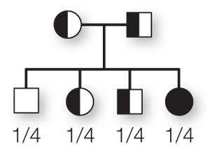
The couple has a 1 in 4 chance of producing a child who is homozygous with two normal alleles, a 1 in 2 chance of producing a child who is heterozygous with one normal allele and one mutant allele, and a 1 in 4 chance of producing a child who is homozygous with two mutant alleles. A child with two mutant alleles would suffer severe anemia and would have to receive regular blood transfusions throughout life.
