Chapter 49
RECAP 49.1
Circulatory systems supply exercising muscle cells with oxygen and nutrients. They also take CO2 and heat out of muscles.
A sponge has water channels throughout its tissues, which means that the external medium can circulate close to all of the sponge’s cells, where exchanges of nutrients, oxygen, and wastes take place.
Hemolymph is the extracellular fluid in animals with an open circulatory system. In animals with a closed circulatory system, blood plasma is the extracellular fluid contained in the heart and blood vessels, and interstitial fluid is the extracellular fluid outside the circulatory system. Blood contains cellular elements in addition to plasma.
The fight-
or- flight response involves increasing blood flow to tissues that are necessary for action, such as the skeletal muscles, and decreasing blood flow to tissues that are not necessary for action, such as the gut. A closed circulatory system can increase blood flow by increasing pressure, and it can also direct that blood flow to critical tissues by changing the resistance in the vessels leading to those tissues.
RECAP 49.2
Deoxygenated blood returns from the systemic circulation in veins that converge on the sinus venous that leads to the atrium. From the atrium, blood flows into the single, muscular ventricle. When the ventricle contracts, blood under pressure flows into the elastic bulbus arteriosus. The elastic recoil of the bulbus arteriosus maintains a continuous blood flow through the gills where respiratory gas exchange occurs. The oxygenated blood from the gills flows through the systemic circuit.
The direction of O2 diffusion across the gill membranes depends on the PO2 gradient between the blood and the water flowing across the gills. By taking air into its lungs, the fish can maintain a higher PO2 in its blood than in the surrounding water. As a result, O2 diffuses from the blood to the water, increasing the PO2 in the water surrounding the eggs.
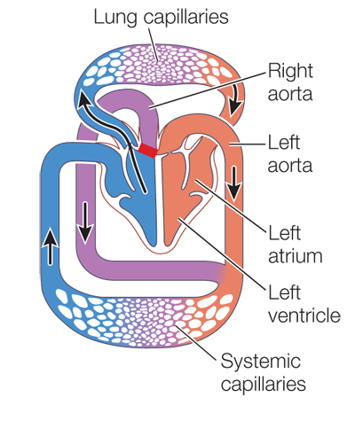
Ectothermic reptiles have a left and a right aorta. Except for the crocodilians, the ventricles are not completely divided and thus blood from the right ventricle can flow either into the right aorta or the pulmonary aorta. When the animal is resting or submerged and not breathing, the resistance in the lung circuit is high and blood from both ventricles flows into the aortas. When the animal is breathing, resistance in the lung circuit goes down, and blood from the right ventricle flows into the lung circuit while blood from the left ventricle flows into the aortas. In crocodilians there is complete separation of the ventricles and the right aorta opens into the right ventricle. But, just where the aortas leave the heart there is a connection between them. When the animal is breathing and resistance in the lung circuit is low, back pressure from the left aorta closes the valve between the right ventricle and right aorta so all blood from the right ventricle flows to the lungs. When resistance in the lung circuit is high, then pressure in the right ventricle is high enough to open the right aortic valve and blood from the right ventricle flows in the right aorta to the systemic circuit.
At birth, when the newborn starts to breathe, the resistance in the pulmonary circuit falls. If the ductus arteriosus does not close, the higher pressure in the left ventricle will pump blood into the pulmonary circuit through the open (patent) ductus arteriosus. This will cause the pulmonary circuit to become congested and the systemic circuit to be deprived of oxygenated blood.
RECAP 49.3
Aortic valve stenosis would decrease the flow of blood into the aorta. Consequences would include decreased pressure in the aorta and major arteries and decreased blood flow to tissues, causing fatigue and shortness of breath. Pressure in the left ventricle would increase, as would its work load. Blood could back up in the pulmonary circuit, causing pulmonary hypertension. Pulmonary valve stenosis would decrease blood flow to the lungs and therefore decrease delivery of oxygenated blood to the left heart, causing fatigue and shortness of breath. The pressure in the right ventricle would increase, as would pressure in the venous return vessels.
Contraction of the myocardium depends on the presence of Ca2+ ions in the sarcoplasm. Increasing Ca2+ in the sarcoplasm prolongs the duration of the Ca2+ pulse resulting from the cardiac action potential. Thus the heart beats more strongly, contractions last longer, and cardiac output increases.
Stenosis of the aortic valve should produce a heart murmur during systole, as that is when pressure in the left ventricle is pushing blood through the aortic valve. Prolapse of the aortic valve should create a heart murmur during diastole, as that is when greater pressure in the aorta is pushing blood back into the ventricle through the prolapsed aortic valve.
RECAP 49.4
The partial pressure of O2 is lower at high altitudes; therefore the athletes experience hypoxia and acclimate to the high altitude environment by producing more red blood cells. As a result, when they compete at lower altitudes, they have greater aerobic capacity.
Blood clotting is a massive event involving large numbers of cells and signals, yet it can be initiated by a small wound that exposes collagen fibers. Platelets that come into contact with those fibers are activated. They become sticky and stick to other platelets, which also become activated. The activated platelets release chemical signals that trigger a long line of clotting reactions, and with each step, the reaction grows. Thus a small event triggers a larger and larger number of events leading to clotting—
a cascade of events. Arterioles are called resistance vessels because of the smooth muscles in their walls that can constrict the vessels and increase their resistance. Changing the resistance of different arterioles allows blood flow to be directed to the tissues and organs that need it the most. Veins are called capacitance vessels because their thin walls with many elastic fibers can stretch to hold larger volumes of blood. When the body is at rest, all of the blood does not have to circulate to serve the body’s needs, so a large proportion of it can be held in reserve in capacitance vessels.
The blood proteins are responsible for the osmotic pressure that draws interstitial fluid back into the capillaries. If the blood proteins are metabolized, the blood osmotic pressure drops and more plasma that leaves the capillaries due to hydrostatic pressure remains in the interstitial spaces. Thus interstitial fluid accumulates in the abdominal cavity and the extremities.
RECAP 49.5
Figure 49.19 shows that a drop in blood flow to a tissue due to a fall in MAP results in autoregulatory responses that decrease the resistance to flow in that tissue. Any decreased resistance in the systemic circuit will cause a drop in MAP, further depriving that tissue of O2 and nutrients and increasing the autoregulatory response. With this positive feedback loop, the autoregulatory response can worsen the problem that initiated it.
The sympathetic division of the ANS stimulates vasoconstriction in tissues not essential for a fight-
or- flight response, such as the digestive system and the skin. This increase in total peripheral resistance results in a rise in MAP. The sympathetic innervation of the heart causes it to beat faster and more strongly, also contributing to a rise in MAP. The parasympathetic division of the ANS has opposite effects, slowing the heart and decreasing MAP. - Page A-51
Baroreceptors are firing at a rate midpoint in their range when blood pressure is normal. Therefore they have the potential to either decrease or increase their firing rate, depending on whether MAP falls or rises. They therefore contribute to regulatory responses to both rises and falls in MAP.
Autoregulatory mechanisms operate at the local level to dilate or constrict arterioles in response to local changes in the chemical environment. These changes in arteriole diameter affect the amount of blood reaching local tissues. As these changes occur, they have an effect on the body’s blood pressure, which is detected by baroreceptors that elicit neural and hormonal signals that cause widespread cardiovascular changes throughout the body.
WORK WITH THE DATA, P. 1055
Comparing mutant mice not CsA treated, but treated with or without RNAi, the left ventricular wall thickness (LVWT) of the controls was 0.96 ± 0.12 and that of the RNAi-
treated mice was 0.70 ± .088. A t-test for the probability that these two mean LVWTs should be the same gives P = 0.0025, indicating that they are significantly different: the RNAi- treated animals had significantly lower LVWT. Comparing mutant mice that did receive CsA but did or did not receive RNAi treatment, the LVWT of the control mice was 1.66 ± 0.22 and that of the RNAi mice was 0.85 ± .06. A t-test calculates P < 0.001, indicating that these two mean LVWTs are significantly different.
FIGURE QUESTIONS
Figure 49.4 If these curves were for the right ventricle, the volume curve would be the same, but the pressure curves during systole would be lower.
Figure 49.9 The breadth of the ventricular muscle action potential is important because it determines the duration of systole and therefore the time available for the emptying of the heart. More complete emptying means greater stroke volume.
Figure 49.10 An occasional block of the AV node could result in a lengthening of the interval between the P and R waves (the PR interval) on the ECG and possibly a missed beat. A long-
APPLY WHAT YOU’VE LEARNED
Blood from the right ventricle is blocked from exiting the right aorta by the surgery. Blood from the right ventricle can pass only through the pulmonary artery. This change converted the crocodilian heart into a heart similar to that of a bird, because a bird heart obligates the blood to go from the right ventricle to the lungs with no opportunity for bypass. In a bird, blood goes from the right ventricle to the lungs to the left atrium to the left ventricle to the body and then back to the right atrium and right ventricle again. In a crocodilian, the blood can go from the right ventricle to either the lungs or the body.
When an alligator is diving and spending time underwater, it is not actively breathing. In these instances (which can extend for long periods of time), the animal does not need to expend energy to pump blood through the lungs. An adaptive advantage is the ability to conserve energy by not pumping blood to the lungs when there is no benefit to be gained from doing so.
If bypassing the pulmonary circuit conserves energy in these animals, an indication might be that growth rates of control animals would have been higher than those of experimental animals, since control animals would have more energy available to divert to growth. However, if we require a P value < 0.01 to conclude that there is a significant difference between the control and experimental groups for both sedentary and exercised animals, there are no significant differences in body mass or body length. This indicates that growth rates between control and experimental animals were similar, suggesting that the pulmonary bypass capability confers no adaptive advantage in terms of growth of juvenile alligators, even when they are stressed by exercise. Therefore these data alone do not allow us to say that the shunt affords a significant conservation of energy related to growth.
An investigator could compare the duration of breath-
hold dives in experimental and control animals. This indicator would provide a measure of the time animals can spend not breathing, which would be an advantage in their ability to survive underwater for extended periods of time. Animals with greater dive times could evade predators more easily and also gain greater predatory advantages themselves by their ability to lie in wait for prey for extended times underwater. A second indicator could be the amount of food consumed and waste produced by experimental and control animals under otherwise identical conditions. This indicator could be used to compute calories burned and would support the hypothesis if experimental animals burned more calories than control animals, since experimental animals would need more energy to go about their daily activities.
A third indicator would be the number of eggs produced by female alligators in experimental and control groups. To support the hypothesis, the results from this study would have to show that experimental females produce fewer eggs over a defined time period than control females. The quantity of eggs is related to the amount of energy the animals have available to put toward reproduction, and loss of the shunt may cause experimental animals to have less energy to expend in producing eggs.