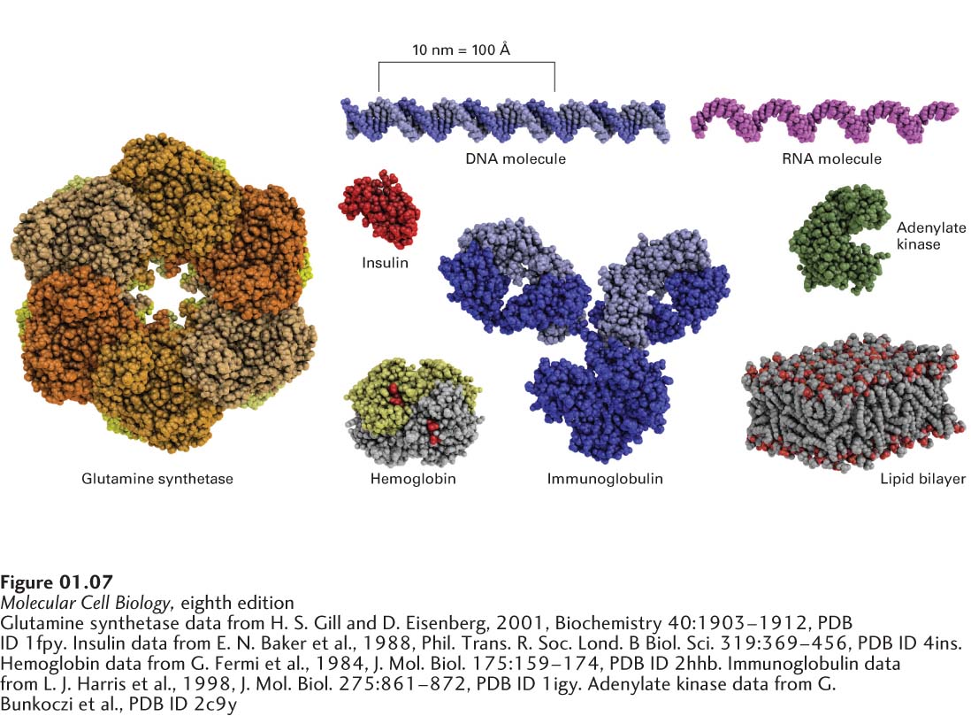
image
FIGURE 1-7 Models of some representative proteins drawn to a common scale and compared with a small portion of a lipid bilayer, a DNA molecule, and an RNA molecule. Each protein has a defined three-dimensional shape held together by numerous chemical bonds. The illustrated proteins include enzymes (glutamine synthetase and adenylate kinase), an antibody (immunoglobulin), a hormone (insulin), and the blood’s oxygen carrier (hemoglobin).
[Glutamine synthetase data from H. S. Gill and D. Eisenberg, 2001, Biochemistry 40:1903–1912, PDB ID 1fpy. Insulin data from E. N. Baker et al., 1988, Phil. Trans. R. Soc. Lond. B Biol. Sci. 319:369–456, PDB ID 4ins. Hemoglobin data from G. Fermi et al., 1984, J. Mol. Biol. 175:159–174, PDB ID 2hhb. Immunoglobulin data from L. J. Harris et al., 1998, J. Mol. Biol. 275:861–872, PDB ID 1igy. Adenylate kinase data from G. Bunkoczi et al., PDB ID 2c9y.]