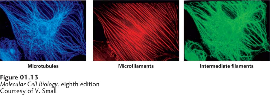
image
FIGURE 1-13 The three types of cytoskeletal filaments have characteristic distributions within mammalian cells. Three views of the same cell. A cultured fibroblast was permeabilized and then treated with three different antibody preparations. Each antibody binds specifically to the protein monomers forming one type of filament and is chemically linked to a differently colored fluorescent dye (green, blue, or red). Visualization of the stained cell in a fluorescence microscope reveals the locations of filaments bound to a particular dye-antibody preparation. In this case, microtubules are stained blue; microfilaments, red; and intermediate filaments, green. All three fiber systems contribute to the shape and movements of cells.
[Courtesy of V. Small.]