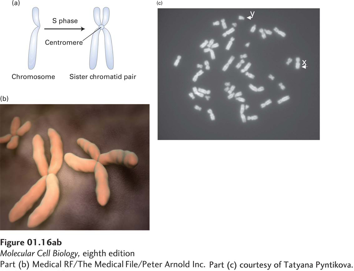
image
FIGURE 1-16 Individual chromosomes can be seen in cells during cell division (mitosis). (a) During the S phase of the cell cycle (see Figure 1-21) chromosomes are duplicated, and the daughter “sister chromatids,” each with a complete copy of the chromosomal DNA, remain attached at the centromere. (b) During the actual cell division process (mitosis), the chromosomal DNA becomes highly compacted, and the pairs of sister chromatids can be seen in the electron microscope, as depicted here. (c) Light-microscope image of a chromosomal spread from a cultured human male lymphoid cell arrested in the metaphase stage of mitosis by treatment with the microtubule-depolymerizing drug colcemid. There is a single copy of the duplicated X and Y chromosomes and two copies of each of the others.
[Part (b) Medical RF/The Medical File/Peter Arnold Inc. Part (c) courtesy of Tatyana Pyntikova.]