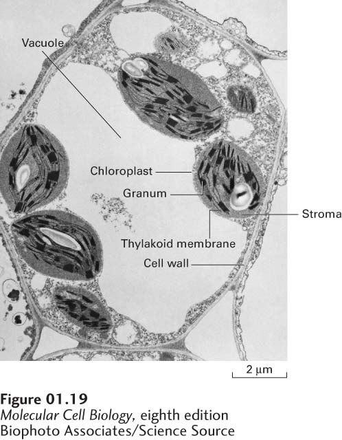
image
FIGURE 1-19 Electron micrograph of a thin section of a leaf cell. In this cell, a single large vacuole occupies much of the cell volume. Parts of five chloroplasts and the cell wall are also visible. Note the internal subcompartments in the chloroplasts.
[Biophoto Associates/Science Source.]