image
FIGURE 10-4 Human hnRNP A1 protein can cycle in and out of the nucleus, but human hnRNP C protein cannot. Cultured HeLa cells and Xenopus cells were fused by treatment with polyethylene glycol, producing heterokaryons containing nuclei from each cell type. These hybrid cells were treated with cycloheximide immediately after fusion to prevent protein synthesis. After 2 hours, the cells were fixed and stained with fluorescent-labeled antibodies specific for human hnRNP C and A1 proteins. These antibodies do not bind to the homologous Xenopus proteins. (a) A fixed preparation viewed by phase-contrast microscopy includes unfused HeLa cells (arrowhead) and Xenopus cells (dotted arrow), as well as fused heterokaryons (solid arrow). In the heterokaryon in this micrograph, the round HeLa-cell nucleus is to the right of the oval-shaped Xenopus nucleus. (b, c) When the same preparation was viewed by fluorescence microscopy, the stained hnRNP C protein appeared green and the stained hnRNP A1 protein appeared red. Note that the unfused Xenopus cell on the left is unstained, confirming that the antibodies are specific for the human proteins. In the heterokaryon, hnRNP C protein appears only in the HeLa-cell nucleus (b), whereas the A1 protein appears in both the HeLa-cell nucleus and the Xenopus nucleus (c). Since protein synthesis was blocked after cell fusion, some of the human hnRNP A1 protein must have left the HeLa-cell nucleus, moved through the cytoplasm, and entered the Xenopus nucleus in the heterokaryon.
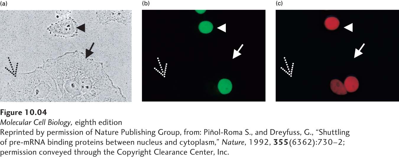
[Reprinted by permission of Nature Publishing Group, from: Piñol-Roma S., and Dreyfuss, G., “Shuttling of pre-mRNA binding proteins between nucleus and cytoplasm,” Nature, 1992, 355(6362):730–2; permission conveyed through the Copyright Clearance Center, Inc.]