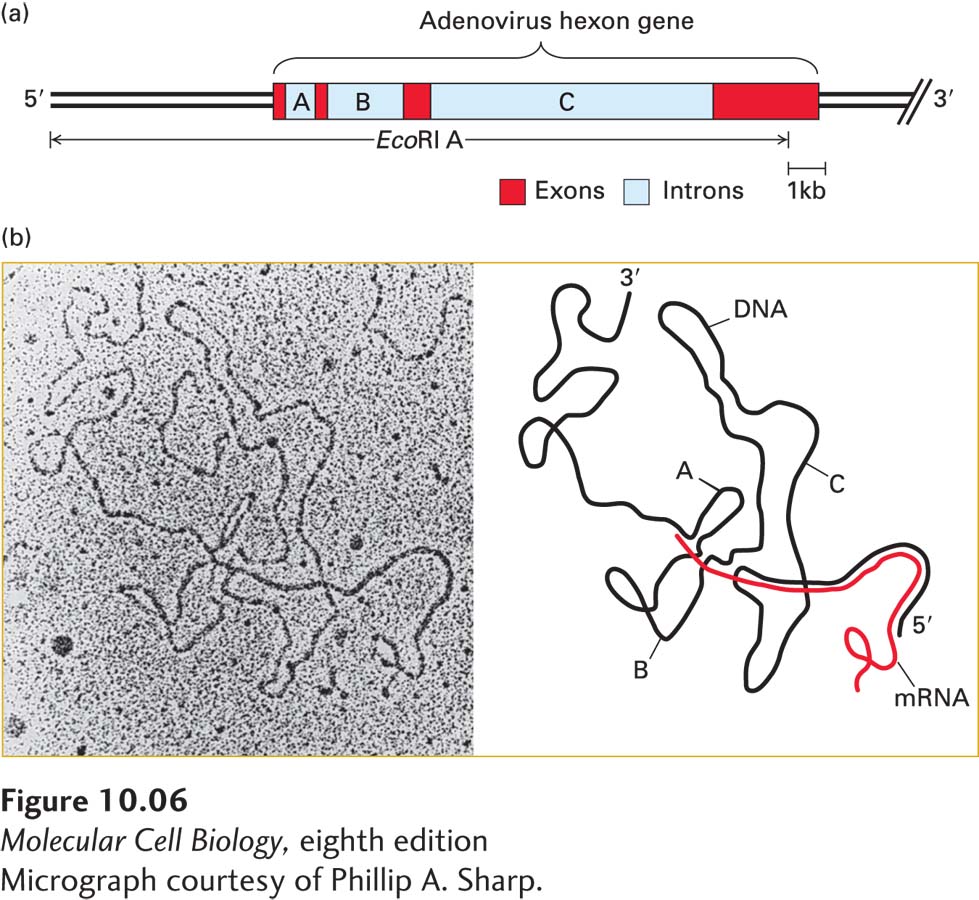
image
EXPERIMENTAL FIGURE 10-6 Electron microscopy of mRNA–template DNA hybrids shows that introns are spliced out during pre-mRNA processing. (a) Diagram of the EcoRI A fragment of adenovirus DNA, which extends from the left end of the genome to just before the end of the final exon of the hexon gene. The hexon gene consists of three short exons and one long (~3.5 kb) exon separated by three introns of ~1, 2.5, and 9 kb. (b) Electron micrograph (left) and schematic drawing (right) of a hybrid between an EcoRI A DNA fragment and a hexon mRNA. The loops marked A, B, and C correspond to the introns indicated in (a). Since these intron sequences in the viral genomic DNA are not present in the mature hexon mRNA, they loop out between the exon sequences that hybridize to their complementary sequences in the mRNA.
[Micrograph courtesy of Phillip A. Sharp.]