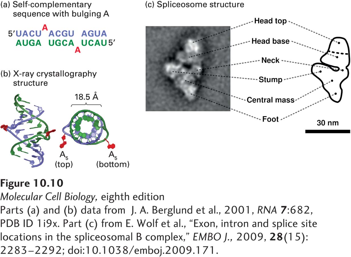
FIGURE 10- 10 Structures of a bulged A in an RNA- RNA helix and an intermediate in the splicing process. (a) Diagram of RNA duplex used for determining the structure of a bulged A. Bulged As at position 5 (red) are excluded from duplex RNA- RNA hybrid formed by complementary bases (blue and green). (b) X- ray crystallography of the structure showed that the bulged A residues extend from the side of an A- form RNA- RNA helix. The phosphate backbone of one strand is shown in green and that of the other strand in blue. The structure on the right is turned 90 degrees for a view down the axis of the helix. (c) 40 Å resolution structure of a spliceosomal splicing intermediate containing U2, U4, U5, and U6 snRNPs, determined by cryoelectron microscopy and image reconstruction. The U4/U6/U5 tri- snRNP complex has a structure similar to the triangular body of this complex below the neck, suggesting that these snRNPs are at the bottom of the structure shown here and that the head is composed largely of U2 snRNP. See H. Stark and R. Luhrmann, 2006, Annu. Rev. Biophys. Biomol. Struct. 35:435.
[Parts (a) and (b) data from J. A. Berglund et al., 2001, RNA 7:682, PDB ID 1i9x. Part (c) from E. Wolf et al., “Exon, intron and splice site locations in the spliceosomal B complex,” EMBO J., 2009, 28(15):2283– 2292; doi:10.1038/emboj.2009.171.]
[Leave] [Close]