image
FIGURE 10-25 Formation of heterogeneous ribonucleoprotein particles (hnRNPs) and export of mRNPs from the nucleus. (a) Model of a single chromatin transcription loop and assembly of Balbiani ring (BR) mRNP in Chironomus tentans. Nascent RNA transcripts produced from the template DNA rapidly associate with proteins, forming hnRNPs. The gradual increase in the size of the hnRNPs reflects the increasing length of RNA transcripts at greater distances from the transcription start site. The model was reconstructed from electron micrographs of serial thin sections of salivary gland cells. (b) Schematic diagram of the biogenesis of hnRNPs. Following processing of the pre-mRNA, the resulting ribonucleoprotein particle is referred to as an mRNP. (c) Model for the transport of BR mRNPs through the nuclear pore complex (NPC) based on electron microscopic studies. Note that the curved mRNPs appear to uncoil as they pass through NPCs. As the mRNA enters the cytoplasm, it rapidly associates with ribosomes, indicating that the 5′ end passes through the NPC first. Parts (b) and (c), see B. Daneholt, 1997, Cell 88:585. See also B. Daneholt, 2001, Proc. Natl. Acad. Sci. USA 98:7012.
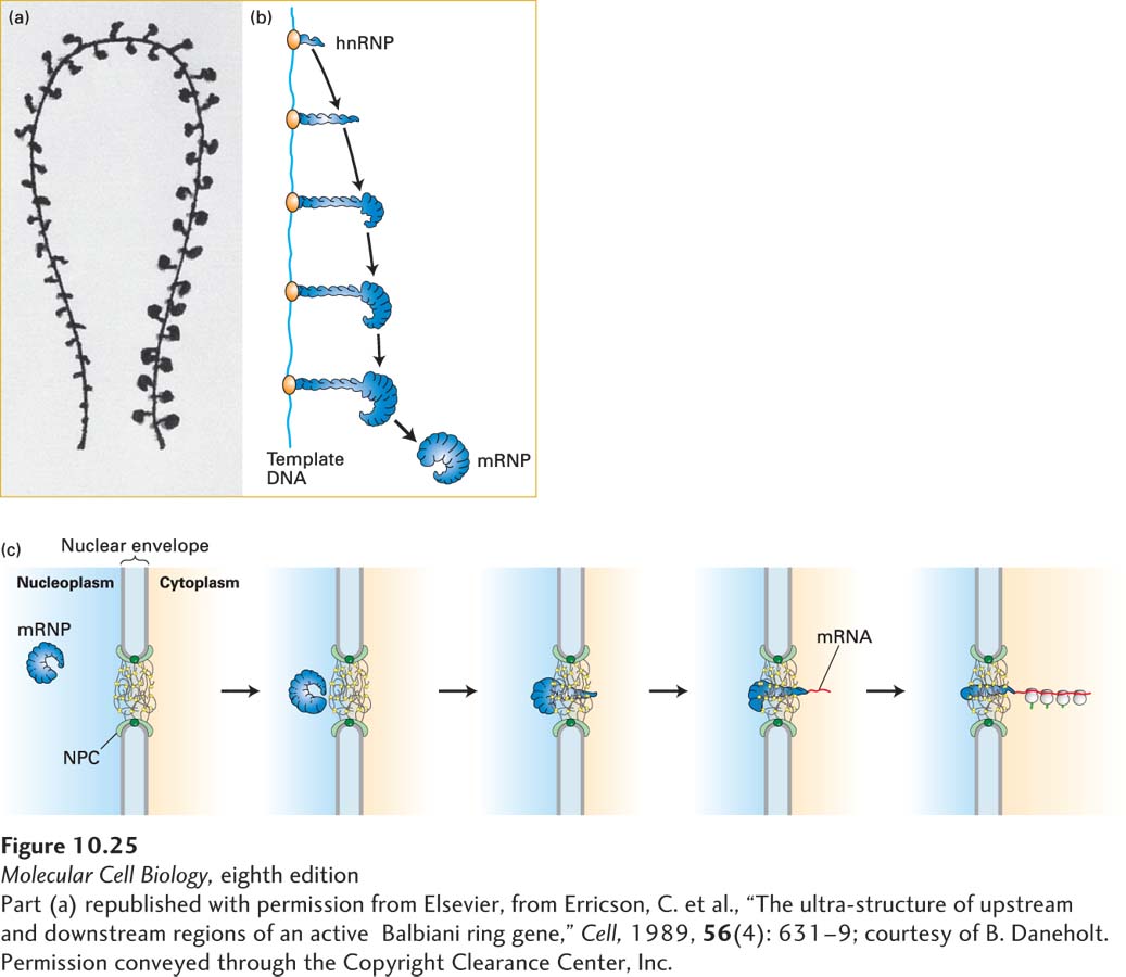
[Part (a) republished with permission from Elsevier, from Erricson, C. et al., “The ultrastructure of upstream and downstream regions of an active Balbiani ring gene,” Cell, 1989, 56(4): 631–9; courtesy of B. Daneholt. Permission conveyed through the Copyright Clearance Center, Inc.]