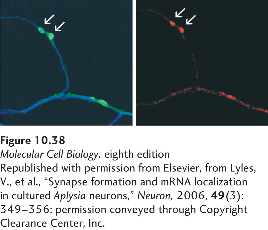
image
EXPERIMENTAL FIGURE 10-38 A specific neuronal mRNA localizes to synapses. Sensory neurons from the sea slug Aplysia californica were cultured with target motor neurons so that processes from the sensory neurons formed synapses with processes from the motor neurons. The micrograph at the left shows motor neuron processes visualized with a blue fluorescent dye. GFP-VAMP (green) was expressed in sensory neurons and marks the location of synapses formed between sensory and motor neuron processes (arrows). The micrograph at the right shows red fluorescence from in situ hybridization of an antisensorin mRNA probe. Sensorin is a neurotransmitter expressed by the sensory neuron only; sensory neuron processes are not otherwise visualized in this preparation, but they lie adjacent to the motor neuron processes. The in situ hybridization results indicate that sensorin mRNA is localized to synapses.
[Republished with permission from Elsevier, from Lyles, V., et al., “Synapse formation and mRNA localization in cultured Aplysia neurons,” Neuron, 2006, 49(3):349–356; permission conveyed through Copyright Clearance Center, Inc.]