image
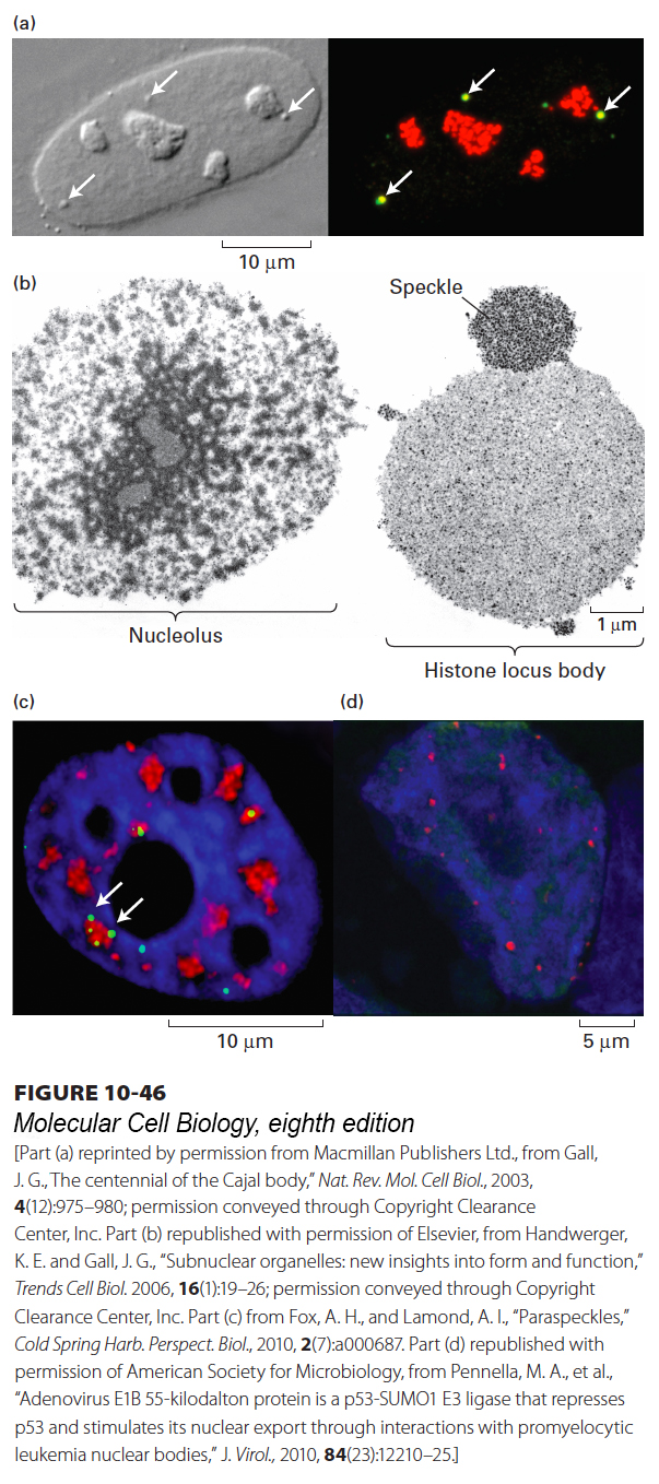
FIGURE 10-46 Examples of nuclear bodies. (a) Cajal bodies and nucleoli in a HeLa cell nucleus. A DIC microscopic image (left) shows four nucleoli and three Cajal bodies (arrowheads). When the same nucleus is immunostained (right) with antibodies against coilin (green) and fibrillarin (red), the three Cajal bodies appear yellow because they stain with both antibodies. The nucleoli stain only for fibrillarin, which is the methyl transferase for 2′-O-methylation of rRNA in the nucleoli and snRNAs in the Cajal bodies. (b) Transmission electron micrograph of nuclear bodies in a single Xenopus oocyte nucleus. Only a portion of the extraordinarily large oocyte nucleus is shown. Histone locus bodies are sites of histone mRNA transcription and processing. They are larger in oocytes, which produce prodigious amounts of histone mRNAs, than in most vertebrate cells. A speckle is a region of concentrated splicing factors. (c) HeLa cell stained with DAPI (blue); antibody to SC35 (red), a splicing factor stored in speckles; and antibody to PSPC1, a protein found in nuclear bodies called paraspeckles (white arrows) because they are most often observed close to speckles. (d) PML nuclear bodies in the nucleus of an H1299 cell (a lung carcinoma cell line). DNA was stained with DAPI (blue) and PML nuclear bodies were immunostained with antibody to the major protein in these bodies, PML.
[Part (a) reprinted by permission from Macmillan Publishers Ltd., from Gall, J. G., “The centennial of the Cajal body,” Nat. Rev. Mol. Cell Biol., 2003, 4(12):975–980; permission conveyed through Copyright Clearance Center, Inc. Part (b) republished with permission of Elsevier, from Handwerger, K. E. and Gall, J. G., “Subnuclear organelles: new insights into form and function,” Trends Cell Biol. 2006, 16(1):19–26; permission conveyed through Copyright Clearance Center, Inc. Part (c) from Fox, A. H., and Lamond, A. I., “Paraspeckles,” Cold Spring Harb. Perspect. Biol., 2010, 2(7):a000687. Part (d) republished with permission of American Society for Microbiology, from Pennella, M. A., et al., “Adenovirus E1B 55-kilodalton protein is a p53-SUMO1 E3 ligase that represses p53 and stimulates its nuclear export through interactions with promyelocytic leukemia nuclear bodies,” J. Virol., 2010, 84(23):12210–25.]