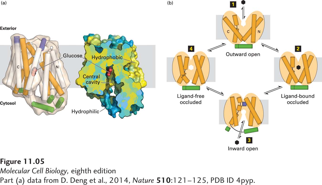
FIGURE 11-5The human GLUT1 uniporter transports glucose across cellular membranes. (a) Structural model (side view) of the full-length human GLUT1 protein in an inward-open conformation. The transporter consists of 12 transmembrane α-helical segments, which are organized into amino-terminal and carboxy-terminal domains, each of which consists of a pair of three transmembrane α helices. The corresponding transmembrane segments in one set of the four three-helix repeats are colored orange in the model on the left. The amino-terminal and carboxy-terminal domains are connected by intracellular and extracellular α helices, which are colored green and purple, respectively. A section of a cut-open view of the surface electrostatic potential highlights the central cavity that transports glucose (red) across the membrane. The colors represent the hydrophobicity of the amino acids, with hydrophobic in yellow and hydrophilic in blue. (b) A working model for GLUT1. In this alternating access model, the outward-open conformation of GLUT1 binds glucose (step 1) and moves to a ligand-bound occluded conformation (step 2) before changing to its inward-open conformation (step 3) when it delivers glucose to the cytoplasm, then moves through a ligand-free occluded conformation (step 4) before beginning another round of glucose transport from outside to inside the cell. If the concentration of glucose is higher inside the cell than outside, the cycle will work in reverse (step 4 → step 1), resulting in net movement of glucose out of the cell. The actual conformational changes are probably smaller than those depicted here.
[Part (a) data from D. Deng et al., 2014, Nature510:121–125, PDB ID 4pyp.]