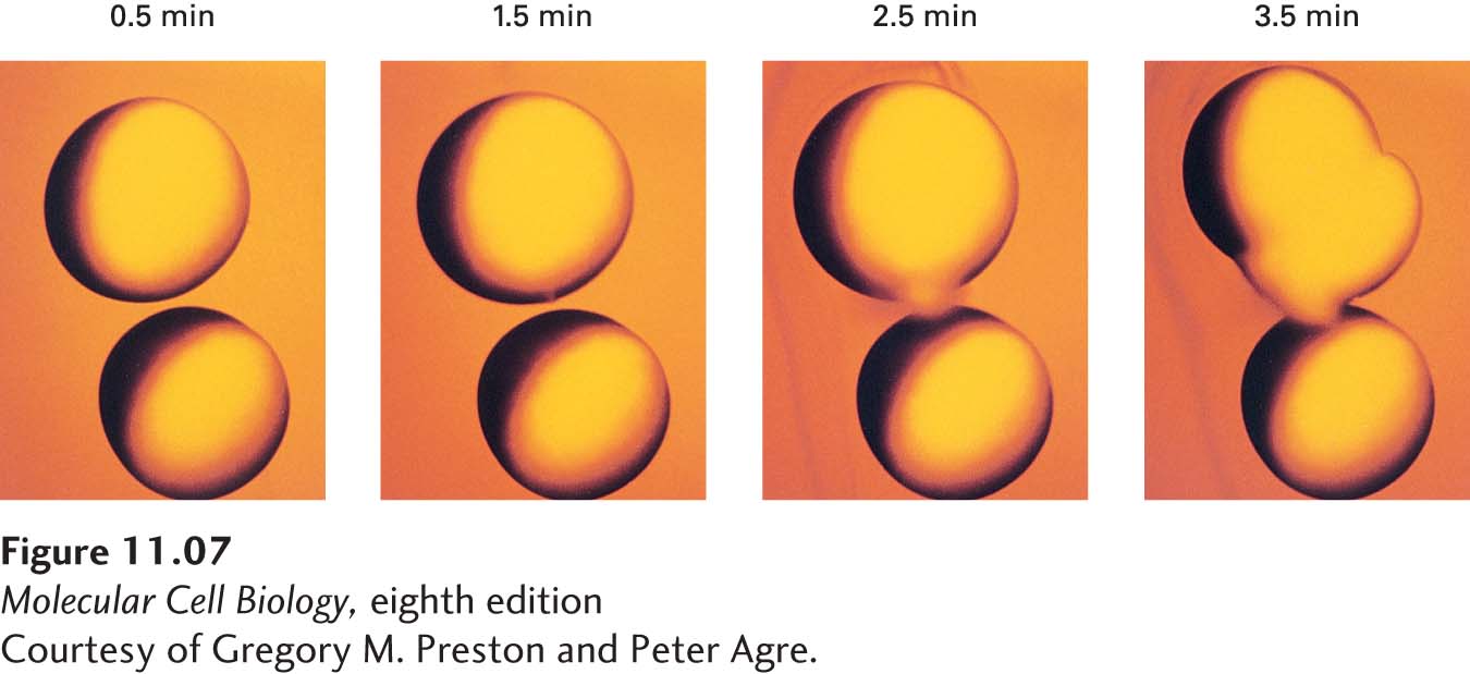
image
EXPERIMENTAL FIGURE 11-7 Expression of aquaporin by frog oocytes increases their permeability to water. Frog oocytes, which normally are impermeable to water and do not express an aquaporin protein, were microinjected with mRNA encoding aquaporin. These photographs show control oocytes (bottom cell in each panel) and microinjected oocytes (top cell in each panel) at the indicated times after transfer from an isotonic salt solution (0.1 M) to a hypotonic salt solution (0.035 M). The volume of the control oocytes remained unchanged because they are not very permeable to water. In contrast, the microinjected oocytes expressing aquaporin swelled and then burst because of an osmotic influx of water, indicating that aquaporin is a water-channel protein. See L. S. King, D. Kozono, and P. Agre, 2004, Nat. Rev. Mol. Cell Biol. 5:687–698.
[Courtesy of Gregory M. Preston and Peter Agre.]