image
FIGURE 11-11 Structure of the catalytic subunit of the muscle Ca21 ATPase. (a) Ca2+-binding sites in the E1 state (left), with two bound calcium ions, and the low-affinity E2 state (right), without bound ions. Side chains of key amino acids are white, and the oxygen atoms on the glutamate and aspartate side chains are red. In the high-affinity E1 conformation, Ca2+ ions bind at two sites between helices 4, 5, 6, and 8 inside the membrane. One site is formed out of negatively charged oxygen atoms from glutamate and aspartate side chains and from water molecules (not shown), and the other is formed out of side-and main-chain oxygen atoms. Seven oxygen atoms surround the Ca2+ ion in both sites. (b) Three-dimensional model of the protein in the E1 state based on the structure determined by x-ray crystallography. There are 10 transmembrane α helices, four of which (purple) contain residues that participate in Ca2+ binding. The cytosolic segment forms three domains: the nucleotide-binding domain (N, blue), the phosphorylation domain (P, green), and the actuator domain (A, beige), which connects two of the membrane-spanning helices. (c) Models of the pump in the E1 state (left) and in the E2 state (right). Note the differences between the E1 and E2 states in the conformations of the N and A domains. Movements of these domains power the conformational changes of the membrane-spanning α helices (purple) that constitute the Ca2+-binding sites, converting them from a conformation in which the Ca2+-binding sites are accessible from the cytosolic face (E1 state) to one in which the now loosely bound Ca2+ ions gain access to the exoplasmic face (E2 state).
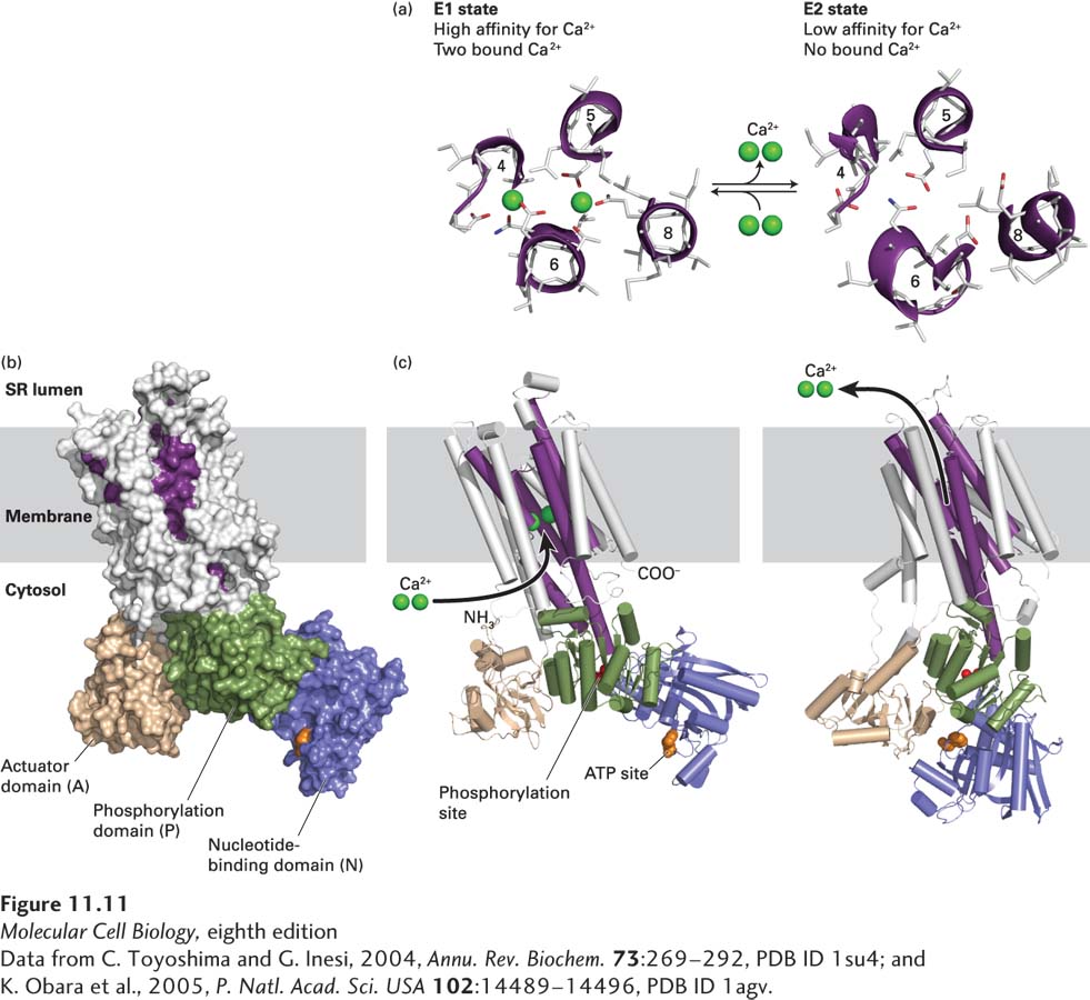
[Data from C. Toyoshima and G. Inesi, 2004, Annu. Rev. Biochem. 73:269–292, PDB ID 1su4; and K. Obara et al., 2005, P. Natl. Acad. Sci. USA 102:14489–14496, PDB ID 1agv.]