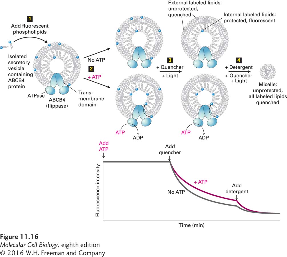
image
EXPERIMENTAL FIGURE 11-16 An in vitro fluorescence-quenching assay revealed the phospholipid flippase activity of ABCB4. A homogeneous population of secretory vesicles containing ABCB4 protein was obtained by introducing the cDNA encoding mammalian ABCB4 into a temperature-sensitive yeast sec mutant such that ABCB4 was localized to intracellular endoplasmic reticulum vesicles in its normal orientation and with the cytosolic face of the vesicles facing outward (see Figure 14-4). Step 1: When synthetic phospholipids containing a fluorescently modified head group (blue) were added to the medium surrounding the purified vesicles, they were incorporated primarily into the outer, cytosolic leaflets of the vesicles. Step 2: If ABCB4 acted as a flippase, then on addition of ATP to the medium, a small fraction of the outward-facing labeled phospholipids would be flipped to the inside leaflet. Step 3: Flipping was detected by adding a non-membrane-permeating quenching compound called dithionite to the medium. Dithionite reacts with the fluorescent head groups, destroying their ability to fluoresce (gray). In the presence of the quencher, only labeled phospholipids in the protected environment of the inner leaflet will fluoresce. Subsequent to the addition of the quenching agent, the total fluorescence decreases with time until it plateaus at the point at which all external fluorescence is quenched and only the internal phospholipid fluorescence can be detected. The observation of greater fluorescence (less quenching) in the presence of ATP than in its absence indicates that ABCB4 has flipped some of the labeled phospholipid to the inside leaflet. Not shown here are “control” vesicles isolated from cells that did not express ABCB4 and that exhibited no flippase activity. Step 4: Addition of detergent to the vesicles generates micelles and makes all fluorescent lipids accessible to the quenching agent, lowering the fluorescence to baseline values. See S. Ruetz and P. Gros, 1994, Cell 77:1071.