image
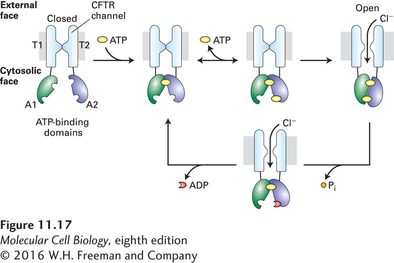
FIGURE 11-17 Structure and function of the cystic fibrosis transmembrane regulator (CFTR). The regulatory (R) domain (not depicted) must be phosphorylated before ATP is able to power channel opening. Upon phosphorylation, one ATP (yellow circle) becomes tightly bound to the A1 domain (green). Binding of a second ATP to the A2 domain (blue) is followed by formation of a tight intramolecular A1–A2 heterodimer and slow channel opening. The relatively stable open state becomes destabilized by hydrolysis of the ATP bound at A2 to ADP (red crescent) and Pi. The ensuing disruption of the tight A1–A2 dimer interface leads to channel closure. T = transmembrane domain; A = cytosolic ATP-binding domain. See D. C. Gadsby et al., 2006, Nature 440:477.