image
FIGURE 11-20 Structure of a resting K+ channel from the bacterium Streptomyces lividans. All K+ channels are tetramers comprising four identical subunits, each containing two conserved membrane-spanning α helices, called by convention S5 and S6, and a shorter P, or pore, segment. (a) One of the subunits, viewed from the side, with key structural features indicated. (b–d) The complete tetrameric channel viewed from the side (b and c) and from the top, or extracellular, end (d). The P segments (green) are located near the exoplasmic surface and connect the S5 and S6 α helices [yellow in (a), yellow and lavender in (b-d)]; they consist of a nonhelical “turret,” which lines the upper part of the pore; a short α helix; and an extended loop that protrudes into the narrowest part of the pore and forms the selectivity filter. This filter allows K+ (purple spheres), but not other ions, to pass. Below the filter is the central cavity, or vestibule, lined by the inner, or S6, α helices. The subunits in gated K+ channels, which open and close in response to specific stimuli, contain additional transmembrane helices not shown here; these channels are discussed in Chapter 22.
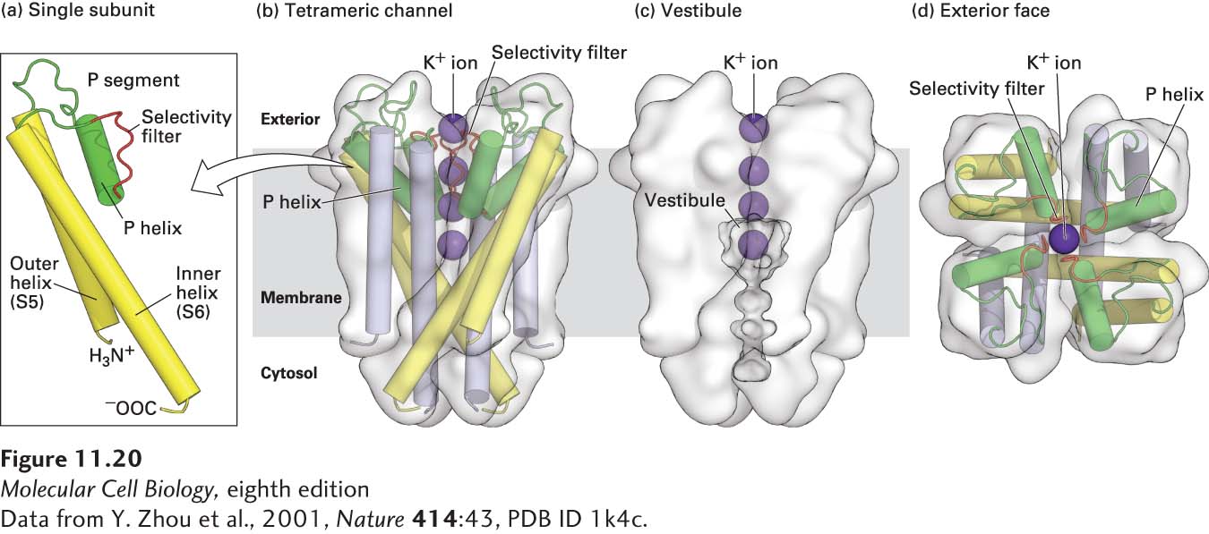
[Data from Y. Zhou et al., 2001, Nature 414:43, PDB ID 1k4c.]