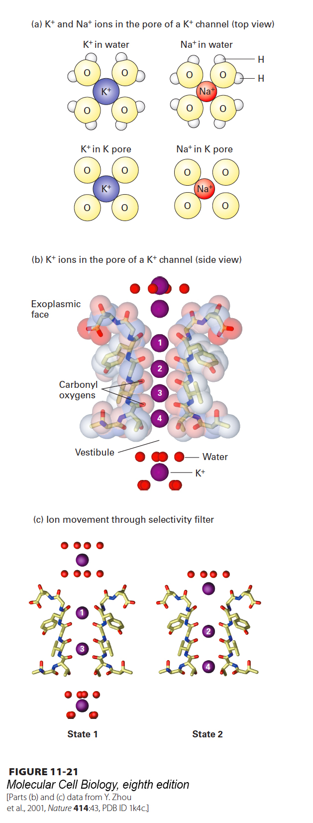
image
FIGURE 11-21 Mechanism of ion selectivity and transport in resting K+ channels. (a) Schematic diagrams of K+ and Na+ ions hydrated in solution and in the pore of a K+ channel. As K+ ions pass through the selectivity filter, they lose their bound water molecules and become bound instead to eight backbone carbonyl oxygens (four of which are shown) that are part of the conserved amino acid sequence in the channel-lining selectivity filter loop of each P segment. The smaller Na+ ions, with their tighter shell of water molecules, cannot perfectly bind to the channel oxygen atoms and therefore pass through the channel only rarely. (b) High-resolution electron density map obtained from x-ray crystallography showing K+ ions (purple spheres) passing through the selectivity filter. Only two of the diagonally opposed channel subunits are shown. Within the selectivity filter, each unhydrated K+ ion interacts with eight carbonyl oxygen atoms (red sticks) lining the channel, two from each of the four subunits, as if to mimic the eight waters of hydration. (c) Interpretation of the electron density map, showing the two alternating states by which K+ ions move through the channel. Ion positions are numbered top to bottom from the exoplasmic side of the channel inward. In state 1, one sees a hydrated K+ ion with its eight bound water molecules, K+ ions at positions 1 and 3 within the selectivity filter, and a fully hydrated K+ ion within the vestibule. During K+ movement, each ion in state 1 moves one step inward, forming state 2. Thus in state 2, the K+ ion on the exoplasmic side of the channel has lost four of its eight waters, the ion at position 1 in state 1 has moved to position 2, and the ion at position 3 in state 1 has moved to position 4. In going from state 2 to state 1, the K+ at position 4 moves into the vestibule and picks up eight water molecules, while another hydrated K+ ion moves into the channel opening, and the other K+ ions move down one step. Note that K+ ions are shown here moving from the exoplasmic side of the channel to the cytosolic side because that is the normal direction of movement in bacteria. In animal cells, the direction of K+ movement is typically the reverse—from inside to outside. See C. Armstrong, 1998, Science 280:56.
[Parts (b) and (c) data from Y. Zhou et al., 2001, Nature 414:43, PDB ID 1k4c.]