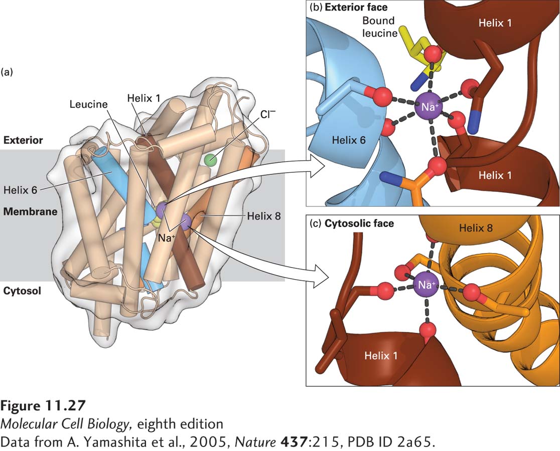
FIGURE 11- 27 Three- dimensional structure of the two- Na+/one- leucine symporter from the bacterium Aquifex aeolicus. (a) The bound L-leucine, two Na+ ions, and a Cl− ion are shown in yellow, purple, and green, respectively. The three membrane- spanning α helices that bind the Na+ or the leucine are colored brown, blue, and orange. (b, c) Binding of the two Na+ ions to carbonyl main- chain or carboxyl side- chain oxygen atoms (red) that are part of helices 1 (brown), 6 (blue), or 8 (orange). It is important that one of the Na+ ions is also bound to the carboxyl group of the transported leucine (part b). See H. Krishnamurthy et al., 2009, Nature 459:347– 355 for details on the structure and function of this and related Na+-linked symporters.
[Data from A. Yamashita et al., 2005, Nature 437:215, PDB ID 2a65.]
[Leave] [Close]