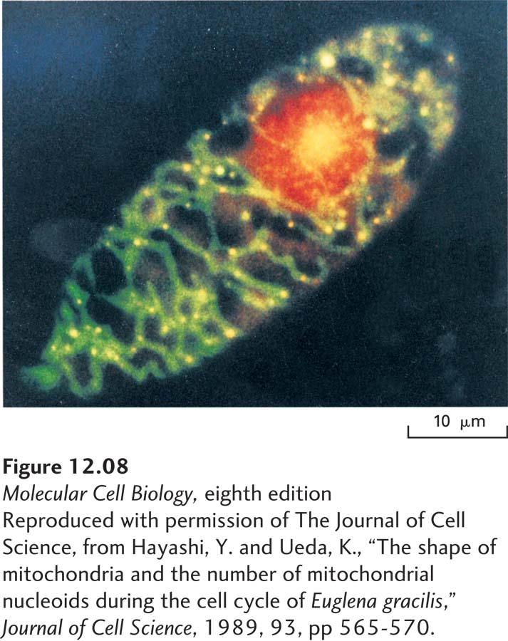
image
EXPERIMENTAL FIGURE 12-8 Dual staining reveals the multiple mitochondrial DNA molecules in a growing Euglena gracilis cell. Cells were treated with a mixture of two dyes: ethidium bromide, which binds to DNA and emits a red fluorescence, and DiOC6, which is incorporated specifically into mitochondria and emits a green fluorescence. Thus the nucleus emits a red fluorescence, and areas rich in mitochondrial DNA fluoresce yellow—a combination of red DNA and green mitochondrial fluorescence.
[Reproduced with permission of The Journal of Cell Science, from Hayashi, Y. and Ueda, K., “The shape of mitochondria and the number of mitochondrial nucleoids during the cell cycle of Euglena gracilis,” Journal of Cell Science, 1989, 93, pp 565-570.]