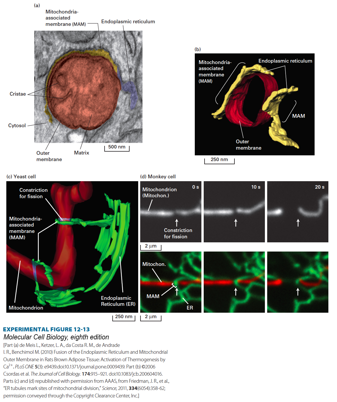
EXPERIMENTAL FIGURE 12- 13 Specialized regions of the endoplasmic reticulum called mitochondria- associated membranes (MAMs) directly contact mitochondria and influence mitochondrial shape, function, and sites of fission. (a) Transmission electron microscopic (EM) image of a section through rat brown adipose (fat) tissue. The lumen of the endoplasmic reticulum (ER) is false colored to show a MAM (yellow) and the non- MAM, bulk ER (blue). The MAM is closely apposed to the outer mitochondrial membrane. (b) Three- dimensional model of a segment of a mitochondrion (red, only outer membrane shown) and the adjacent MAM (yellow) determined from a line of cultured avian lymphoma cells using EM tomography (assembly of a three- dimensional image from consecutive individual sections). (c) A three- dimensional model of a mitochondrion (red) and adjacent MAMs (green) from a yeast cell using EM tomography. The two MAM domains are derived from ER tubules that in some cases can wrap around the mitochondrion, in the top case forming a clamp- like structure that appears to constrict the mitochondrion in preparation for fission. (d) Live cell fluorescence microscopic images of a Cos- 7 monkey cell, showing a mitochondrion (white in the top panels, same mitochondrion in red in the bottom panels) and MAM (green in bottom panels), taken from a single field of view at 10- second intervals. The arrow points to the site of constriction and fission on the mitochondrion and to the MAM at the constriction/fission site. The MAM directs constriction and subsequent DRP1- mediated fission at this site. To visualize the mitochondria and ER, the Cos- 7 cells were transfected with cDNA vectors encoding two fluorescent proteins that specifically accumulate in either the mitochondrion (red fluorescence) or the ER (green fluorescence).
[Part (a) de Meis L., Ketzer, L. A., da Costa R. M., de Andrade I. R., Benchimol M. (2010) Fusion of the Endoplasmic Reticulum and Mitochondrial Outer Membrane in Rats Brown Adipose Tissue: Activation of Thermogenesis by Ca2+. PLoS ONE 5(3): e9439.doi:10.1371/journal.pone.0009439. Part (b) ©2006 Csordas et al. The Journal of Cell Biology. 174:915– 921. doi:10.1083/jcb.200604016. Parts (c) and (d) republished with permission from AAAS, from Friedman, J. R., et al., “ER tubules mark sites of mitochondrial division,” Science, 2011, 334(6054):358- 62; permission conveyed through the Copyright Clearance Center, Inc.]
[Leave] [Close]