image
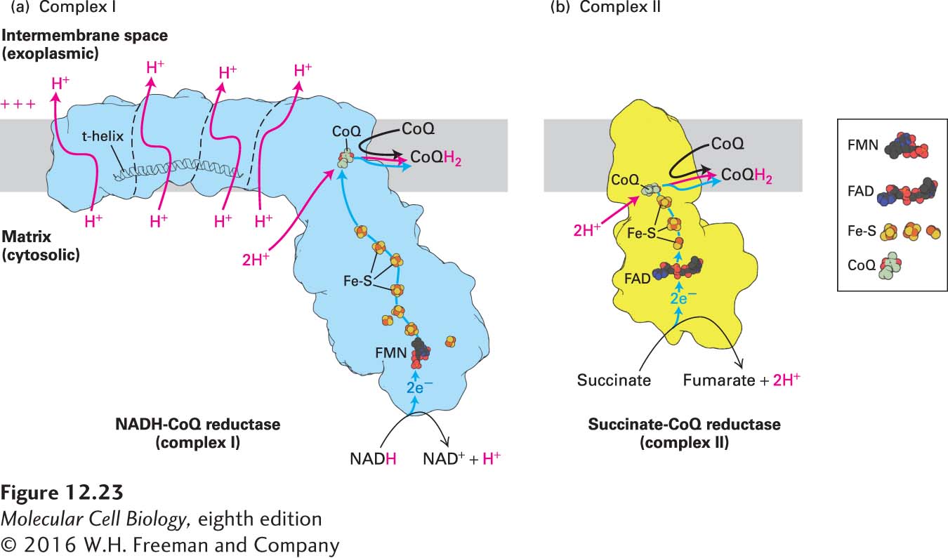
FIGURE 12-23 Electron and proton transport through complexes I and II. (a) Model of complex I based on its three-dimensional structure. The outline of the shape of the core complex I from the yeast Y. lipolytica, as determined by x-ray crystallography, is shown in light blue, and the borders separating several distinct structural subunits are indicated by thin dashed black lines. From NADH, electrons flow first to a flavin mononucleotide (FMN) and then, via iron-sulfur clusters (Fe-S, red and yellow balls), to CoQ, to which two protons from the matrix bind (red arrow) to form CoQH2. Conformational changes due to the electron flow, which may be induced by changes in the charge of the CoQ and may include a piston-like horizontal movement of the t-helix, drive proton pumping through the transmembrane subunits from the matrix to the intramembrane space (red arrows). (b) Model of complex II based on its three-dimensional structure. Electrons flow through complex II from succinate to CoQ via FAD/FADH2 and iron-sulfur clusters (Fe-S), and from complex II to complex III via CoQ/CoQH2. Electrons released during oxidation of succinate to fumarate in complex II are used to reduce CoQ to CoQH2 without translocating additional protons.
[Part (a) data from V. Zickermann et al., 2015, Science 347:44–49, PDB ID 3m9s. Part (b) data from F. Sun et al., 2005, Cell 121:1043–1057, PDB ID 1zoy.]