image
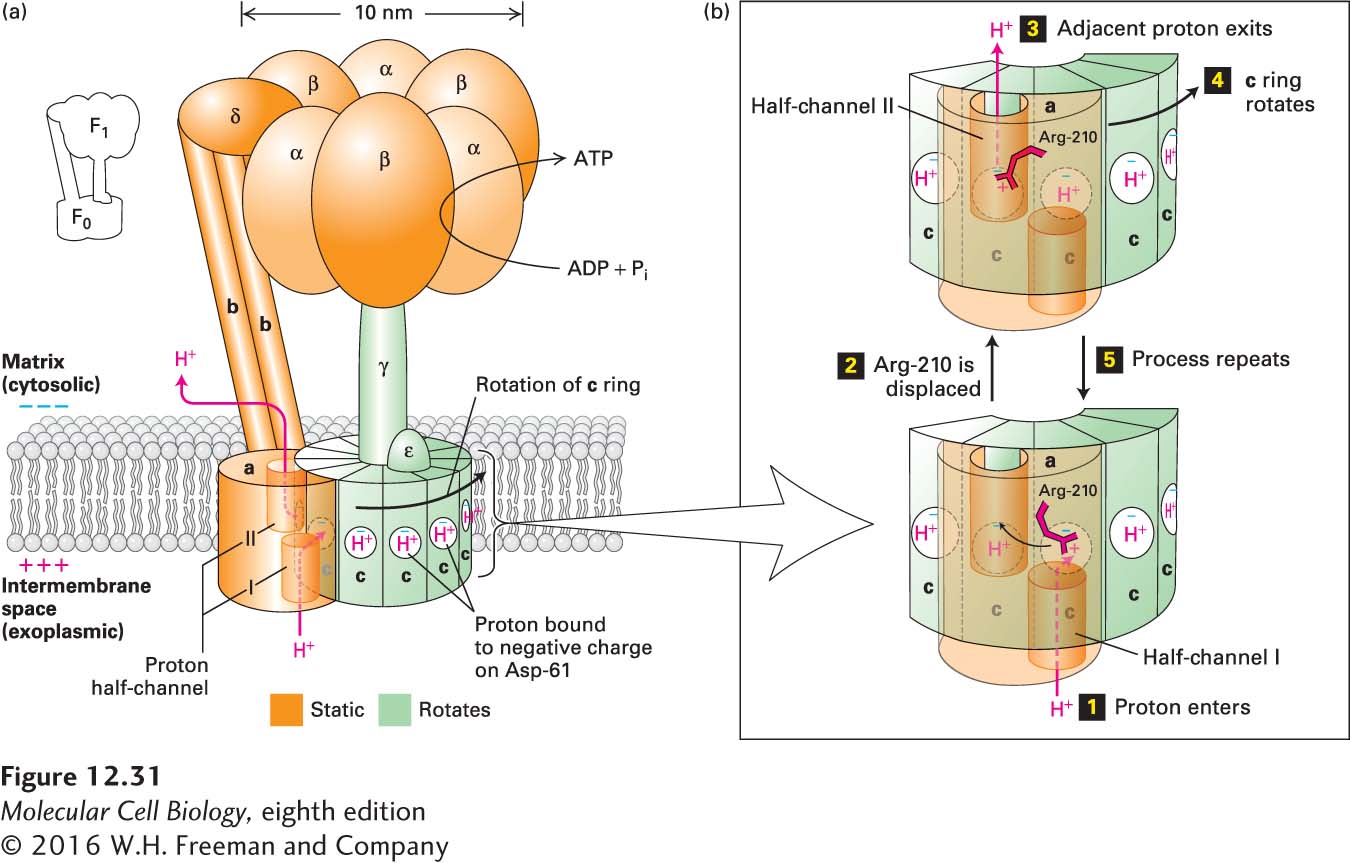
FIGURE 12-31 Structure of ATP synthase (the F0F1 complex) in the bacterial plasma membrane and mechanism of proton translocation across the membrane. (a) The F0 membrane-embedded subcomplex of ATP synthase is built of three integral membrane proteins: one copy of a, two copies of b, and an average of ten copies of c arranged in a ring in the plane of the membrane. Two proton half-channels near the interfaces of subunit a with the c subunits mediate proton movement across the membrane (proton path is indicated by red arrows). Half-channel I allows protons to move one at a time from the exoplasmic medium (equivalent to intermembrane space in mitochondria) to the negatively charged side chain of Asp-61 in the center of a c subunit near the middle of the membrane. The proton-binding site in each c subunit is represented as a white circle with a blue “−” representing the negative charge on the side chain of Asp-61. Half-channel II permits protons to move from the Asp-61 of an adjacent c subunit into the cytosolic medium. The detailed structure of the c ring and a portion of the adjacent a subunit is shown in Figure 12-34. The F1 subcomplex of ATP synthase contains three copies each of subunits α and β, which form a hexamer resting atop the single rod-shaped γ subunit, which is inserted into the c ring of F0. The ε subunit is rigidly attached to the γ subunit and also to several of the c subunits. The δ subunit permanently links one of the α subunits in the F1 subcomplex to the b subunit of F0. Thus the F0 a and b subunits and the F1 δ subunit and (αβ)3 hexamer form a rigid structure (orange) anchored in the membrane. During proton flow, the c ring and the attached F1 ε and γ subunits rotate as a unit (green), causing conformational changes in the F1 β subunits, leading to ATP synthesis. (b) Potential mechanism of proton translocation. Step 1: A proton from the exoplasmic space enters half-channel I and moves toward the “empty” (unprotonated) Asp-61 proton-binding site. The negative charge (blue “−”) on the unprotonated side chain Asp-61 is balanced, in part, by a positive charge on the side chain of Arg-210 (red “+”). Step 2: The proton fills the empty proton-binding site and simultaneously displaces the positively charged Arg-210 side chain, which swings over to the filled proton-binding site on the adjacent c subunit (curved arrow). As a consequence, the proton bound at that adjacent site is displaced. Step 3: The displaced adjacent proton moves through half-channel II and is released into the cytosolic space, leaving an empty proton-binding site on Asp-61. Step 4: Counterclockwise rotation of the entire c ring moves the “empty” c subunit over half-channel I. Step 5: The process is repeated. See M. J. Schnitzer, 2001, Nature 410:878; P. D. Boyer, 1999, Nature 402:247; and C. von Ballmoos, A. Wiedenmann, and P. Dimroth, 2009, Annu. Rev. Biochem. 78:649.